Article by Dr. Thomas Burnell and Bethany Turner
Questions by Dr. Josh Sturgeon
Next Lesson - Artery and Vein Disease
Abstract
- Heart failure is the inability of the heart to meet the demands of the body.
- It can be divided into heart failure with reduced ejection fraction (e.g. caused by arrhythmias) or preserved ejection fraction (e.g. caused by hypertension).
- The symptoms of right sided heart failure are based around blood building up in the systemic circulation (e.g. peripheral oedema), and those of left sided heart failure are to do with blood building up in the pulmonary circulation (E.g. orthopnoea).
- Treatment of heart failure is focused on reducing symptoms.
Core
Heart failure is the inability of the heart to meet the body’s demands. It is a clinical syndrome of reduced cardiac output (CO), tissue hypoperfusion, increased pulmonary pressure and tissue congestion.
There are many causes of heart failure, including:
- Ischaemic heart disease – this causes myocardial damage which causes myocardial dysfunction, reducing the cardiac output.
- Hypertension – increases the afterload on the ventricle. This causes the ventricles to undergo hypertrophy in order to generate more force to pump blood out against the higher pressure.
- The thickened myocardium results in heart being is less efficient at pumping blood as there is reduced space in the ventricle reducing the amount of filling.
- Aortic stenosis – increases the afterload on the ventricle.
- Other valvular/myocardial structural diseases
- Cardiomyopathies – e.g. dilatation or hypertrophy.
- Arrhythmias
- Pericardial disease
- Cor pulmonale – right ventricular heart failure secondary to chronic lung disease, e.g. COPD, pulmonary fibrosis, bronchiectasis.
- If the body’s demands become excessive, requiring a grossly elevated cardiac output e.g. sepsis, severe anaemia, thyrotoxicosis.
A main cause of heart failure is the stroke volume (SV) is reduced. The causes of this can be split into three categories:
- Reduced pre-load – filling of the ventricles, so less blood is pumped out
- Reduced myocardial activity – heart muscle can no longer produce the same force of contraction for a given volume so less blood is pumped out.
- Increased after-load – the heart is pumping against a higher pressure leading to hypertrophy.
Heart failure has two main classifications based on the equation:
Ejection fraction = (Stroke Volume) / (End Diastolic Volume). Normal ejection fraction is >50%, reduced ejection fraction is <40%.
The classifications are:
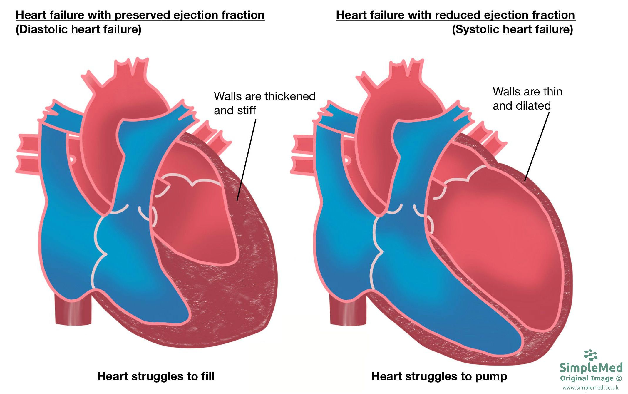
- Heart failure with reduced ejection fraction, HFrEF – where there is systolic dysfunction.
- In HFrEF, there is a problem with heart contraction and therefore the heart cannot pump with enough force, so there is systolic dysfunction.
- Causes include: thin/fibrosed walls, enlarged chambers, abnormal/uncoordinated contraction.
- It is also known as systolic heart failure.
- Heart failure with preserved ejection fraction, HFpEF – where there is diastolic dysfunction.
- In HFpEF, there is a problem with the heart where it does not fill properly, so there is diastolic dysfunction.
- The main cause for diastolic dysfunction is hypertrophy. This is as the chambers become stiff and cannot relax enough to allow adequate filling of the ventricles.

Diagram - The classification of heart failure
SimpleMed original by Bethany Turner
Heart failure can also be classified based on the symptoms that patients experience. The classes are:
- Class I – no symptomatic limitation of physical activity.
- Class II – slight limitation of physical activity. No symptoms at rest.
- Class III – marked limitation of physical activity. No symptoms at rest.
- Class IV – inability to carry out physical activity without symptoms. May have symptoms at rest.
Heart failure one on side of the heart can lead to heart failure on the other side of the heart. When there is both left and right sided heart failure, this is called congestive or biventricular heart failure.
In heart failure the decreased CO causes a decreased BP (BP = CO x TPR). Reduced BP causes:
- Baroreceptors to detect the low BP and stimulate an increase sympathetic drive that leads to:
- Increased heart rate
- Increased peripheral resistance
- Activation of the Renin-Angiotensin-Aldosterone System that causes:
- Increased sodium retention leading to increased water retention (water follows sodium) and this increases the circulating volume
- Anti-Diuretic Hormone release
- Vasoconstriction
- Enhanced sympathetic activity
This is all an attempt to increase the BP but in this reality culminates in an increased after-load and pre-load which puts even more strain on the failing heart.
Signs and Symptoms of Heart Failure
The signs and symptoms of heart failure depend on the side of the heart affected. This determines whether the pulmonary or systemic circulation is predominantly affected.
Right sided heart failure signs and symptoms:
- Fatigue/lethargy
- Breathlessness
- Peripheral pitting oedema
- Raised jugular venous pressure
- Tender, smooth and enlarged liver (hepatomegaly)
Left sided heart failure signs and symptoms:
- Fatigue/lethargy
- Breathlessness
- Pulmonary oedema which can manifest as:
- Orthopnoea
- Paroxysmal nocturnal dyspnoea - attacks of breathlessness at night which wakes the patient from sleep
- Basal pulmonary crackles
- Cardiomegaly (enlarged left ventricle leads to a displaced apex beat on examination)
Investigations for Heart Failure
Full Blood Count – check for anaemia as the symptoms of anaemia are similar to heart failure. Anaemia can worsen heart failure as it puts further strain on the heart to meet the body's demands.
Electrolytes and renal function – chronic kidney failure causes fluid overload as they are not removing the fluid from the body. Fluid overload can cause the symptoms of heart failure.
Brain Natriuretic Peptide (BNP) – BNP is produced by the stretching of ventricles. It is produced to try and reduce BP by increasing sodium loss and therefore increasing water loss.
- An elevated BNP suggests heart failure while a normal BNP with breathlessness usually excludes heart failure.
- It can be used to monitor progression.
ECG – an abnormal ECG with a raised BNP provides evidence for heart failure.
Chest X-Ray – pulmonary oedema can be classically seen on a chest x-ray as batwing shadowing. An enlarged heart can also be seen on a chest x-ray as an increased cardiothoracic ratio (ratio of maximal horizontal cardiac diameter to maximal horizontal thoracic diameter on a chest X-ray).
Echocardiogram – can be used to measure the ejection fraction to help determine the type of heart failure. It can also assess the valve and ventricular function to help determine the causative factor.
Coronary angiography – this technique allows for imaging of the coronary arteries and can be used to determine if they are blocked by an atherosclerotic plaque. If there is a blockage of a coronary artery, the oxygen delivery to the myocardium is reduced, putting strain on the heart. Blockage can lead to ischaemia and acute coronary syndromes.
Management of heart failure depends on whether it is acute or chronic.
Acute is managed in hospital in the following way:
- Must give oxygen.
- Give an IV loop-diuretic, Furosemide. This causes diuresis (water loss) to reduce the volume of fluid in the circulation. This will reduce the after-load and increase the cardiac output.
- Giving a diuretic also helps to reduce oedema.
- Give heparin to reduce the risk of venous clots forming or to prevent propagation of those which have formed.
- The management may require additional ventilator support and IV nitrates.
Chronic heart failure is managed by:
- Correcting the underlying cause:
- E.g. heart transplant, mechanical assist device (e.g. in valve disorders), pacemaker (e.g. in arrhythmia), implanted defibrillator.
- Non-pharmacological management:
- Reduce salt intake, increase aerobic exercise, reduce alcohol intake.
- Pharmacological therapy – this is mainly for symptomatic improvement, to delay the progression of heart failure.
- ACE inhibitors, angiotensin-receptor blockers, β-blockers, spironolactone (aldosterone receptor antagonist), diuretics.
- The aim of pharmacological therapy is to reduce the after-load and increase the CO. This is for symptomatic control of heart failure.
Edited by: Dr. Ben Appleby
- 12041

