By Joshua Bray
Next Lesson - Oral Cavity and Pharynx
Abstract
- The nose has many functions: these include olfaction, filtering, humidifying and warming inspired air, speech and drainage.
- The nasal skeleton is predominantly hyaline cartilage with a proximal bony root.
- The nasal septum is cartilaginous anteriorly and bony posteriorly.
- Septal haematomas, if left untreated, can lead to saddle nose deformity.
- The nasal cavity is lined with olfactory mucosa and respiratory mucosa.
- There are 3 pairs of conchae, which increase the opportunity for air humidification and warming.
- There are 4 pairs of paranasal sinuses: frontal, ethmoidal, maxillary and sphenoidal.
- There is a rich anastomosis of blood vessels in the anterior nasal septum (Kiesselbach’s area), which is a common source of epistaxis.
- The nasal cavity is innervated by the olfactory nerve and trigeminal nerve.
- Rhinitis is inflammation of the nasal mucosa and includes both the common cold and hay fever.
- Viral infection of the nose can spread to the paranasal sinuses, resulting in sinusitis.
- Nasal polyps are fleshy benign growths of the nasal mucosa and are usually bilateral.
- Unilateral nasal discharge in an adult should raise suspicion for nasal cancer.
Core
The nose is part of the respiratory tract situated immediately superior to the hard palate.
Functions of the nose include:
- Olfaction (smell)
- Conduction of inspired air
- Filtration of inspired air
- Humidification and warming of inspired air
- Resonance chamber for speech
- Drainage of secretions from the paranasal sinuses and nasolacrimal ducts
The external nose is the visible portion of the nose which protrudes from the face. Its prominent nature makes it particularly susceptible in facial trauma.
The nose has two external openings called the nares (or nostrils). Via the nares, inspired air immediately enters the vestibule of the nose, which is lined with skin containing sebaceous glands and hairs which assist in trapping unwanted particles from the air.
The skeleton of the nose is predominantly hyaline cartilage with a proximal bony root.
The bony part of the nose consists of the nasal bones, frontal processes of the maxillae, nasal part of the frontal bone and the bony part of the nasal septum.
There are 5 nasal cartilages:
- 2 Lateral Cartilages – situated just inferior to the nasal bones. They are flattened and triangular in shape.
- 2 Alar Cartilages – the ‘squishy’ part of the external nose. These cartilages are mobile and can dilate with contraction of the nasalis muscle.
- 1 Septal Cartilage – the cartilaginous part of the nasal septum.
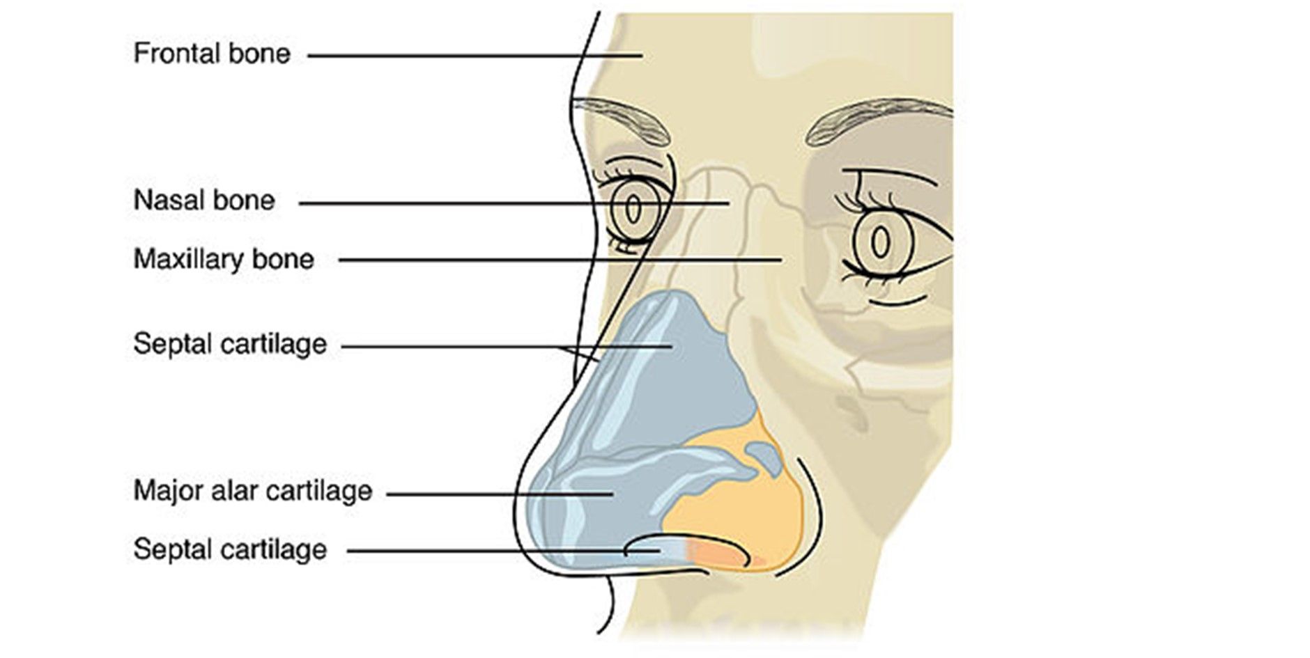
Diagram - The nasal skeleton
Creative commons source by OpenStax College, edited by Joshua Bray [CC BY-SA 4.0 (https://creativecommons.org/licenses/by-sa/4.0)]
The nasal cavity is the internal space of the nose, which extends from the vestibule to the nasopharynx. It is practically horizontal in nature.
The boundaries of the nasal cavity are:
- Roof – nasal bone and cartilages
- Floor – hard palate
- Lateral Walls – maxilla (mainly)
- Medial Wall – nasal septum
The nasal septum separates the left and right nasal cavities. It has 2 parts:
- Cartilaginous (anteriorly)
- Bony (posteriorly) – consisting of the perpendicular portion of the ethmoid bone and the vomer bone
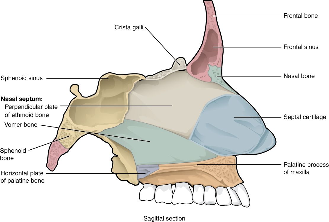
Diagram - The nasal septum
Creative commons source by OpenStax College [CC BY-SA 4.0 (https://creativecommons.org/licenses/by-sa/4.0)]
Trauma to the nose could shear blood vessels in the nasal septum, causing a septal haematoma. This haematoma separates the cartilage from its overlying perichondrium, depriving the cartilage of its blood supply. If left untreated, the septal cartilage undergoes avascular necrosis and remodelling, resulting in a saddle-nose deformity.
The nasal cavity is lined with mucosa and is highly vascular.
The olfactory mucosa lines the roof of the nasal cavity and houses olfactory neurones, which are responsible for our sense of smell. They produce action potentials in response to chemical stimuli (aromas), which are conducted along the olfactory nerve (CN I).
The rest of the nasal cavity is lined with respiratory mucosa. It is composed of pseudostratified columnar epithelium and houses cilia and mucous glands. It acts to warm, humidify and filter inspired air.
The nasal conchae (or turbinates) are bony shelf-like projections from the lateral walls of the nasal cavity. There are 3 pairs of conchae: superior, middle and inferior. Each concha has a respective meatus, which is a gap underneath the curve of the concha. The function of the conchae is to induce turbulent air flow, thereby slowing air flow and increasing the opportunity for humidification and warming.
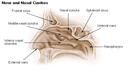
Diagram - The nasal cavity and conchae
Public Domain Source (NIH) [Public domain]
The paranasal sinuses are air-filled extensions of the nasal cavity. They are lined with respiratory mucosa and act to warm and humidify inspired air.
There are 4 pairs of sinuses:
- Frontal – situated between the outer and inner tables of the frontal bone.
- Ethmoidal – otherwise termed ‘ethmoidal air cells’. They are small invaginations of mucosa into the ethmoid bone.
- Maxillary – the largest of the paranasal sinuses, located within the body of the maxilla, just inferior to the orbit.
- Sphenoidal – located within the body of the sphenoid bone.
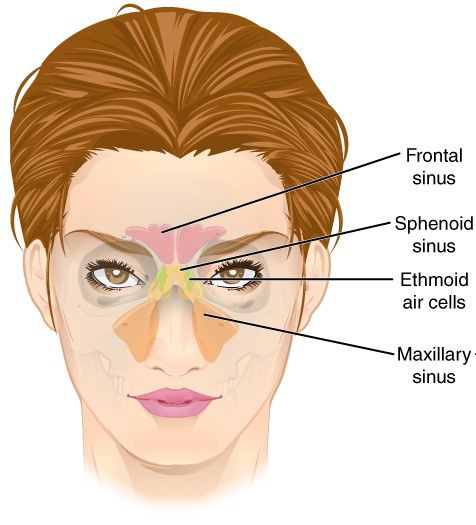
Diagram - Paranasal air sinuses
Creative commons source by OpenStax College [CC BY-SA 4.0 (https://creativecommons.org/licenses/by-sa/4.0)]
The paranasal sinuses have important anatomical relations with:
- Nasal Cavity – the sinuses drain directly into the nasal cavity.
- Orbit – the maxillary sinus is situated immediately inferior to the orbit. Fractures to the floor of the orbit (‘orbital blow-out’ fracture) can cause the contents of the orbit to prolapse into the maxillary sinus.
- Anterior Cranial Fossa – the frontal, ethmoidal and sphenoidal sinuses are in close proximity to the anterior cranial fossa.
- Roots of Upper Teeth – these can sometimes project into the maxillary sinuses.
The nasal cavity is supplied by branches of the ophthalmic, maxillary, and facial arteries:
- Anterior ethmoidal artery (ophthalmic a.)
- Posterior ethmoidal artery (ophthalmic a.)
- Sphenopalatine artery (ophthalmic a.)
- Greater palatine artery (maxillary a.)
- Superior labial artery (facial a.)
These arteries anastomose in the anterior nasal septum. This anastomosis is named Kiesselbach’s plexus or Little’s area and is a common source of epistaxis.
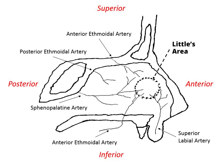
Diagram - Arterial supply of the nasal cavity
Creative commons source by Mbuchko3 [CC BY-SA 4.0 (https://creativecommons.org/licenses/by-sa/4.0)]
Venous blood is drained via the sphenopalatine, facial, and ophthalmic veins. There is also a communication with the cavernous sinus, hence the nose is part of the ‘danger triangle’ of the face, where infections can potentially spread intracranially.
Epistaxis (a nosebleed) is often a minor and self-limiting problem. However, persistent bleeds can be serious and lead to hypovolaemia.
The most common cause of epistaxis is minor trauma to the nose. However, predisposing factors include coagulopathies, connective tissue disorders and malignancy.
Epistaxis can be anterior or posterior in origin:
- Anterior – this is the most common source, arising from Kiesselbach’s area.
- Posterior – arising from the sphenopalatine artery. These bleeds are less common and more difficult to treat.
The management steps for epistaxis are as follows (in this order):
- First Aid – pinch the alar cartilages for at least 15 minutes with the head tilted forward.
- Cautery – in an attempt to seal off the bleeding vessel.
- Anterior Packing – using nasal tampons.
- Posterior Packing – using a balloon catheter.
- Surgical Intervention (last resort) – either embolization or ligation of the vessel.
Special sensory innervation (olfaction) is supplied via the olfactory nerve (CN I).
The somatosensory innervation of the nasal cavity is via branches of the trigeminal nerve (CN V):
- Anterosuperiorly – via ophthalmic division (CN Va)
- Posteroinferiorly – via the maxillary division (CN Vb)
Conditions Affecting the Nose and Sinuses
Rhinitis is an umbrella term for inflammation of the nasal mucosa. It encompasses both the common cold (coryza) and allergic rhinitis (hay fever). Typical symptoms include nasal irritation, sneezing, nasal discharge and congestion.
The common cold is caused by a range of respiratory viruses, including rhinoviruses, adenoviruses and coronaviruses. It is transmitted via droplet spread and is highly contagious, however disease is often mild and self-limiting. In addition to nasal symptoms, individuals may experience fever, fatigue, malaise and sore throat.
Sometimes viral infection can spread from the nose to the paranasal sinuses, resulting in sinusitis. Roughly 2% of cases are complicated by a bacterial infection (with Streptococcus pneumoniae or Haemophilus influenzae). Clinical features include frontal headache, purulent nasal discharge, facial pain and tenderness, and fever. Sinusitis is normally self-limiting, however, bacterial sinusitis can be treated with antibiotics. Bacterial sinusitis is more likely if symptoms are particularly severe at onset, persist after 10 days or get worse after initial improvement.
Hay fever is a manifestation of a Type I hypersensitivity reaction (allergy). B cells recognise allergens and produce IgE, which binds to mast cells and causes them to release histamine, resulting in local inflammation. Common allergens include pollen, dust mites, animal dander and mould. Hay fever also commonly affects the eyes and is associated with other atopic illness such as asthma and eczema. Management is with oral antihistamines and intranasal corticosteroids.
Nasal polyps are fleshy benign growths of the nasal mucosa. Affected individuals may complain of nasal discharge (+/- blood), blocked nose, loss of smell or taste, cough (due to post-nasal drip) and snoring. Nasal polyps are usually bilateral. Unilateral nasal discharge should raise suspicion for nasal cancer.
In children, another important differential for nasal discharge is a foreign body.
Reviewed by: Dr. Thomas Burnell
- 4480

