Next lesson - Introduction to DNA
Core
Cells are the units that make up the human body - each cell has a role within the body for which it is designed and specialised (e.g. protection - keratinized squamous epithelial cells of the skin - or secretion - mucous-secreting columnar goblet cells of the respiratory tract). This article covers the common components of cells in the form of the hypotheical "standard" eukaryotic cell (cells with enveloped nuclei).
An organelle is a component of the cell with a specific function.

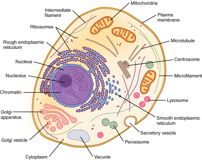
Diagram - The eukaryotic cell, with its various organelles labelled, each with a specific function
Creative commons source by 3.2 The Cytoplasm and Cellular Organelles by Rice University [CC BY-SA 4.0 (https://creativecommons.org/licenses/by-sa/4.0)]
- The main central organelle of the cell.
- Contains DNA, nucleoproteins (nucleic acids bonded to a protein) & RNA.
- Function: stores DNA and co-ordinate the cell’s activities, such as growth, metabolism, protein synthesis and cell division.
- Transmission electron microscope images of the nucleus reveal that DNA is found in two forms. Heterochromatin (condensed DNA (chromatin) -> APPEARS DARK- heterochromatin has no active RNA synthesis as it is condensed) and euchromatin (uncondensed DNA -> APPEARS LIGHT - has active RNA synthesis).
- N.B. Prokaryotic cells will not have a nucleus which is what defines them as prokaryotc.
- Found within the nucleus,
- Function: the site of ribosomal RNA (rRNA) synthesis. The site at which it is combined with proteins to generate incomplete ribosomes. These ribosomes then mature after being exported from the nucleus into the cytoplasm, where they either attach to rough endoplasmic reticulum or become free-floating ribosomes within the cytoplasm.
- A double layered membrane bounding the nucleus.
- Contains nuclear pores - these allow macromolecules to pass through when entering or exiting the nucleus.
- Function: controls what enters and exits the nucleus.
Rough Endoplasmic Reticulum (RER)
- A series of interconnecting membranes, vesicles & cisternae (flattended sacs), continuous throughout the cytoplasm.
- Ribosomes are attached to the outer surface of the membranes, making this ER "rough".
- Function: synthesises proteins destined for lysosomes, the cell membrane or extracellular export.
- Pancreatic cells (and other cells which synthesise substances) have abundant RER to synthesise pancreatic enzymes.
Smooth Endoplasmic Reticulum (SER)
- A series of interconnecting membranes, vesicles and cisternae (less flat compared to RER) that are less extensive throughout the cytoplasm. The lack of embedded ribosomes classifies this ER as smooth.
- Function: lipid biosynthesis and intracellular transport (e.g. steroid production).
- Composed of two rRNA subunits (40s+60s for eukaryotes and 50s +30s for prokaryotes) that wrap around mRNA to begin translation and protein synthesis.
- Function: site of protein synthesis (translation) within the cell.
- Semi-circular shaped stacks of cisternae.
- Vesicles containing proteins bud off from the RER and fuse with the convex forming face of Golgi body.
- The Golgi bodies have polarity - proteins move from the convex (entry point) to concave end of the stack and are modified as they move.
- Function: sort, concentrate, package and modify proteins synthesised in the RER.
- Vesicles containing different proteins leave the maturing, concave face of the Golgi body, destined for lysosome assembly - they then undergo further processing or secretion.
- Membrane bound organelles that contains acid hydrolases at pH 5 (proteases, nucleases, glycosidases, lipases and phosphatases).
- Function: break down excess or worn out organelles and digested viruses or bacteria.
- A membrane bound organelle that contains enzymes that transfer hydrogen atoms from toxins such as alcohol to oxygen to produce hydrogen peroxide.
- The toxins ingested are neutralised and the hydrogen peroxide produced can be neutralised by the enzymes in peroxisomes to produce water and oxygen.
- Function: chemical detoxification and lipid metabolism.

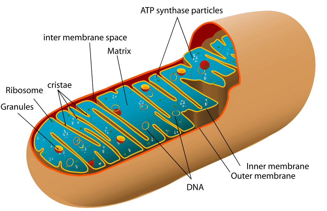
Diagram - The structure of the mitochondria
Creative commons source by Mariana Ruiz Villarreal LadyofHats [CC BY-SA 4.0 (https://creativecommons.org/licenses/by-sa/4.0)]
- A key component of the cell. Particularly abundant in highly active, energy requiring cells.
- Mitochondria have a double membrane with an outer and inner lipid bilayer membrane.
- The inner membrane is larger than the outer membrane and is therefore highly folded to fit within the outer membrane. This folding increases the surface area for ATP generation to occur - these folds are called cristae.
- Contained within the inner membrane lies the matrix where the TCA/Krebs cycle takes place. The matrix contains mitochondrial DNA, ribosomes and soluble enzymes.
- The enzymes in the matrix are responsible for the creation of ATP in the TCA/Krebs cycle and oxidative phosphorylation.
- Cells such as muscle cells and sperm cells have a higher number of mitochondria due to increased requirements for energy in the form of ATP.
- Function: Responsible for the generation of ATP viz. energy, for the cell through aerobic respiration and regulates cellular metabolism

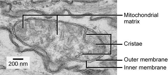
Diagram - Labelled image of a mitochondria under a microscope
Creative commons source by CNX OpenStax [CC BY-SA 4.0 (https://creativecommons.org/licenses/by-sa/4.0)]
- The plasma membrane is composed of phospholipids. These are essentially triacyl glycerides (TAGs) with a phosphate head instead of a fatty acid chain. Phospholipids are amphipathic molecules (have both hydrophobic and hydrophilic regions)
- There are proteins embedded in the plasma membrane. These can be either peripheral proteins (only exist on one side of the membrane) or integral proteins (span the width of the bilayer).
- Cholesterol is present in plasma membranes and controls the fluidity or rigidity of the membrane at various temperatures.
- Take a look at our Biological membranes article for more detail on structure of membranes
- Functions:
- Act as a selectively permeable barrier as it only allows non-polar, small molecules through without the aid of channels.
- Compartmentalises cells into discreet units.
- Communication with other molecules and cells.
- Recognition through signalling molecules, adhesion proteins and immune surveillance.
- Electrical or chemical signal generation in response to a stimuli.
- All cells posses a cytoskeleton which is responsible for maintaining and changing the cell’s shape.
- There are three main structural components of the cytoskeleton: microfilaments, intermediate filaments and microtubules.
- Function:
- Provides structural support for the plasma membrane & cell organelles.
- Provides a means of movement for organelles, plasma membrane and other cytosol constituents around the cell.
- Provides the locomotor mechanism for amoebic movement of cells such as lymphocytes (crawling-like movement) and is the basis of cilia and flagella.
- Provides contractility in cells of specialised tissues (e.g. muscle cells).
- Microfilaments:
- Composed of two strings of actin and measure 5nm in diameter.
- They are associated with ATP allowing for contractility. They can also assemble and dissociate very quickly (dynamic).
- The structure of microvilli of the intestinal cells is maintained by a core of actin filaments.
- Intermediate filaments:
- Unlike microfilaments, they are not dynamic and are 10-12nm in diameter.
- Their function is to form a tough supporting meshwork within the cytoplasm. They are anchored to the plasma membrane at desmosomes (strong intracellular junctions).
- The filaments are commonly found in nerve cells, neuroglial cells and epithelial cells (made out of cytokeratin).
- Microtubules:
- Tubulin subunits polymerise to form the wall of the hollow microtubule.
- They originate from the centrosome and found at sites in the cell where structures are moved - nerve fibres, mitotic spindles, cores of cilia & flagella.
- Attachment proteins can bind to the organelles and move the structures along the microtubules e.g. vesicles moving through the cytoplasm to the plasma membrane.
Edited by: Dr. Ben Appleby and Dr. Marcus Judge
- 11803

