By Dr. Joshua Sturgeon and Dr. Ben Appleby
Next Lesson - Phenotype, Genotype and Inheritance
Abstract
- Mitosis is the method of cell division for somatic cells and involves replication of DNA (replicated chromosomes contain two identical sister chromatids joined by a centromere) followed by one round of division, resulting in 2 diploid daughter cells.
- Meiosis is the method of cell division for germ cells and involves replication of DNA followed by two rounds of division, resulting in 4 haploid gametes.
- Nondisjunction occurs when homologous chromosomes are not equally distributed in the daughter cells after mitosis or meiosis. Nondisjunction can lead to aneuploidy, the cause of conditions such as Down’s Syndrome (trisomy 21).
Core
Chromosome StructureThe term chromosome refers to an unreplicated DNA polymer. However, the popular image of an classical X-shape chromosome is in fact a chromosome which has replicated but may confusingly be referred to as a chromosome (implying not replicated). The replicated chromosome contains two identical DNA polymers, with each polymer (arm of the ‘X’) referred to as a sister chromatid. Maternal and paternal pairs which are not made up of replicated DNA are referred to as a homologous pair.
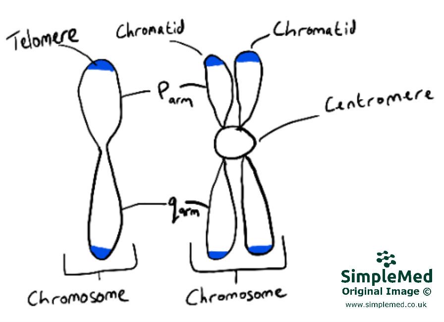
Image: The structure of a chromosome before (left) and after (right) replication of DNA has occurred. Note that the replicated chromosome contains two sister chromatids.
SimpleMed original by Joshua Sturgeon
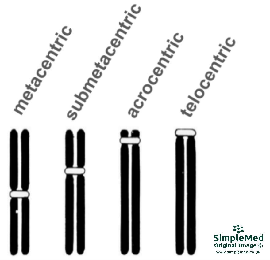
Image: Different centromere locations.
SimpleMed original by Joshua Sturgeon
Chromosomes are grouped according to size and shape:
Image: (Extension material) Chromosome group classifications.
SimpleMed original by Joshua Sturgeon
During interphase, chromosomes do not exist as shown in the previous two figures (i.e. as rods) but instead as loose filaments of uncondensed chromatin. The chromosomes only take on the appearance of rods (condensed chromosomes) during division once the chromatin condenses together in prophase.
Although the chromatin is uncondensed and free the chromosomes still occupy specific spaces known as chromosome territories. (Knowledge of the specific areas occupied is not crucial.)
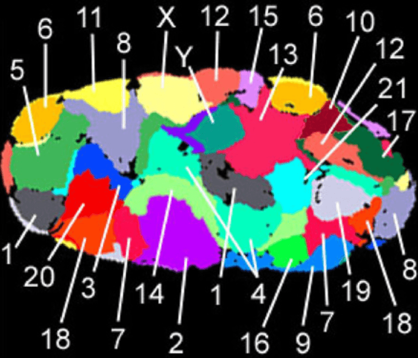
Image: Chromosome territory map.
Creative commons source by Andreas Bolzer, Gregor Kreth, Irina Solovei, Daniela Koehler, Kaan Saracoglu, Christine Fauth, Stefan Müller, Roland Eils, Christoph Cremer, Michael R. Speicher, Thomas Cremer [CC BY 2.5 (https://creativecommons.org/licenses/by/2.5)]
Each chromosome is paired, these pairings are known as homologous chromosomes. Homologous chromosomes contain the same genes, but they may have different variants of the genes (alleles).
Karyotyping is a method of describing the chromosome composition of a patient. The karyotype is recorded in a standard form including chromosome number, sex complement (XX or XY), and any structural changes. Information in a karyotype is separated by a comma without spaces. A karyotype is derived from a metaphase spread which is a visualisation of all the chromosomes from a cell.
46,XX = Normal Female
46,XY = Normal Male
47,XY,+21 = Male with trisomy 21, also known as Down’s syndrome.
Mitosis is the method of cell division for somatic cells. The process results in two identical daughter cells with the same chromosome content as the parent cell. Mitosis is crucial for growth, regeneration and repair in the human body by allowing for new tissue to be produced.
There are 5 phases of mitosis:
Prophase – centrioles and spindle fibres develop; chromatin condenses into condensed chromosomes. (Genetic replication has already occurred during interphase which precedes cell division.)
Prometaphase – nuclear envelope dissolves and spindle fibres attach to the kinetochore (found on the centromere).
Metaphase – replicated chromosomes line up along the equator of the cell.
Anaphase – sister chromatids are split and are pulled to opposite poles of the cell.
Telophase – nuclear envelope reforms, the cytoplasm begins to cleave. The chromosomes have now been split from their replicated sister chromatid.
These phases are followed by cytokinesis, which is the splitting of the cytoplasm which results in the formation of 2 daughter cells.
Mitosis starts with a somatic body cell with 46 chromosomes, organised into 23 pairs. When replication of this DNA occurs, there are still 46 chromosomes, but each one is made up of two chromatids joined by the centromere, making 92 chromatids in this cell. When telophase occurs, it is the centromere that splits, dividing the two chromatids and leaving two daughter cells with 46 chromosomes (each cell has one chromatid, which becomes a chromosome in its own right when separated).
Non-disjunction is the failure of one or more pairs of sister chromatids to separate normally during nuclear division, which results in an abnormal distribution of chromosomes in the daughter cells. In mitosis, this means one of the daughter cells will have 3 copies of one chromosome (trisomy) and one will have only 1 copy (monosomy).
The consequences of mitotic non-disjunction depend on when in fetal development the non-disjunction occurs.
If nondisjunction occurs in the first post-zygotic division (first cell division in a developing embryo making two cells from one) a trisomy cell (with 47 chromosomes) and a monosomy cell (with 45 chromosomes) will be produced. The monosomy cell will be destroyed leaving only the trisomy cell to divide. From this point onward all cells in the body will now be trisomy which may lead to conditions such as Down’s syndrome . This is a non-mosaic karyotype.
If the non-disjunction event happens during a later cell division a slightly different outcome is produced. As before the monosomy cell is destroyed and a trisomy cell is produced. However, by this point multiple successful divisions have occurred meaning there are also surviving diploid cells. In the long term, some cells in the body will be derived from the trisomy cell line, and the rest will be derived from the healthy, normal diploid cell line – this presence of more than one cell line in the body is called mosaicism. This mosaicism can be spread throughout the tissues of the body if division happened earlier, or may be limited to a specific tissue.
Meiosis is a special form of cell division used for production of germ cells. These germ cells have 46 chromosomes, arranged in 23 pairs, same as any other body cell. Meiosis results in the production of four non-identical cells, each with half the chromosome content of the parent cell (a haploid cell).In meiosis, one round of replication is followed by two rounds of division, which can be split into meiosis I and II.
Meiosis I is identical to mitosis, with the cell moving through the same phases with one crucial difference:
In mitosis the sister chromatids of a replicated chromosome are pulled apart in anaphase through the splitting of the centromere e.g. the maternal chromosome 11 pair of sister chromatids has its two replicated arms pulled apart leaving each cell with a single copy of the maternal chromosome. This means that at the end of mitosis, there are two daughter cells with exactly the same DNA.
In meiosis however, during anaphase I (in meiosis I) the homologous pairs (ie the 23 pairs) are pulled apart leaving each cell with a pair of sister chromatids; this means that each cell has 23 chromosomes and 46 chromatids. It also means that genetic information is randomly distributed between the cells with some of the cells after meiosis I carrying paternal genetic information and some carrying maternal genetic information. For example, the maternal chromosome 11 during anaphase I is pulled away from the paternal chromosome 11 (the other half of its homologous pair).
In meiosis II, the sister chromatids are split through division of the centromere, becoming chromosomes in their own right, leaving each daughter cell with 23 chromosomes (which were chromatids a moment ago).
In summary:
- Stem cell begins with 46 chromosomes arranged in 23 pairs.
- The cell undergoes DNA replication to form 46 chromosomes, each with 2 chromatids, meaning 92 chromatids.
- The cell undergoes first division, dividing the 23 pairs, forming two cells each with 23 chomosomes, formed from 46 chromatids.
- The cell undergoes second division, splitting the centromeres, leaving four daughter cells in total, each with 23 chomosomes (made up of 23 chromatids).
Non-disjunction in meiosis occurs in the same way as mitotic non-disjunction. The severity of the consequences of meiotic non-disjunction depends if the non-disjunction event happens in meiosis I or II. If it occurs in meiosis I, after fertilisation, none of the zygotes will be normal, they will either be trisomic or monosomic.
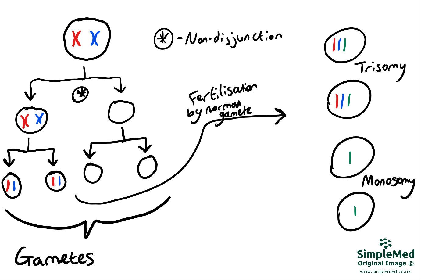
SimpleMed original by Joshua Sturgeon
If non-disjunction happens in meiosis II, after fertilisation, 2 zygotes will be normal, one will be monosomic, and one will be trisomic.
96% of Down’s Syndrome cases are caused by non-disjunction in meiosis.
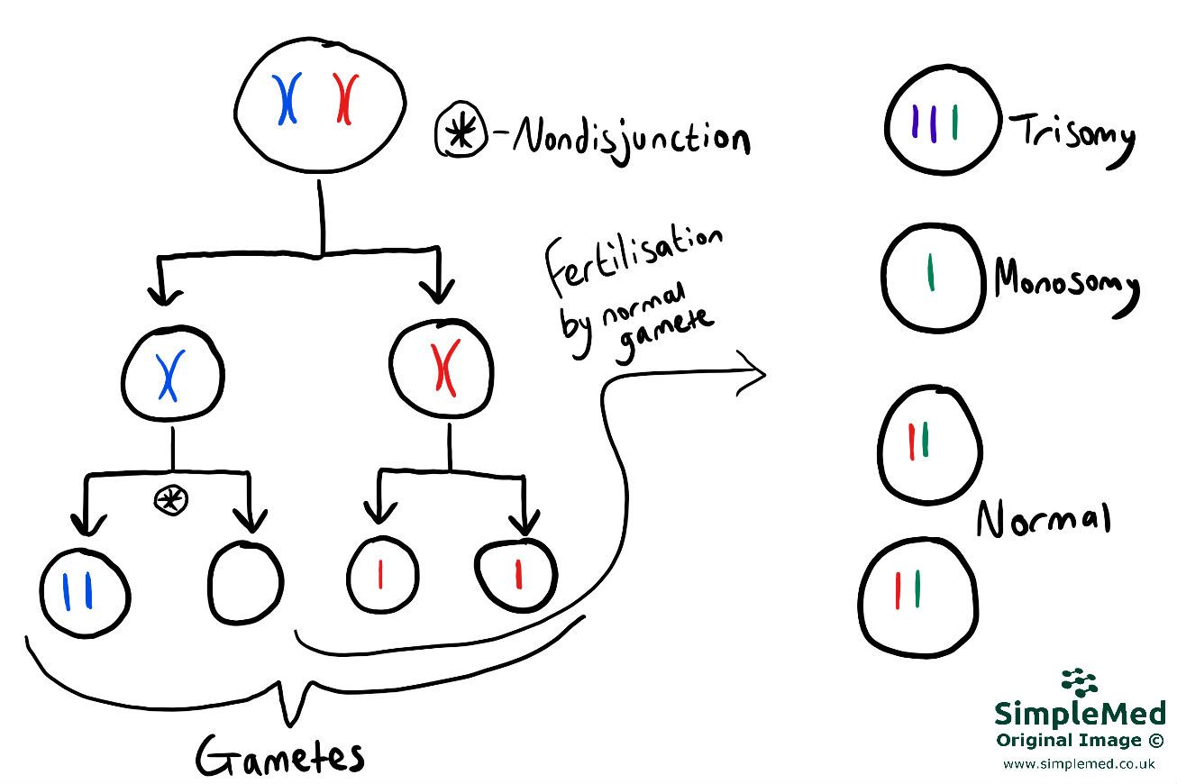
Image: Non-disjunction in Meiosis 2.
The umbrella term for an incorrect number of chromosomes in a cell- i.e. not 46 - is known as aneuploidy. Aneuploidy can cause a number of genetic conditions depending on whether chromosomes are, missing or in excess, and which specific chromosomes are affected.
Conditions caused by an addition of a single chromosome leading to a total of 47.
- Down’s syndrome: 47,XX,+21 – addition of a chromosome 21 – a total of 3 chromosome 21’s. This karyotype would be for a female patient
- Edward’s syndrome: 47,XX,+18 – addition of a chromosome 18. This karyotype would be for a female patient. Patients with this condition unfortunately do not generally survive past infancy.
- Patau’s syndrome: 47,XX,+13 – addition of a chromosome 13. This karyotype would be for a female patient. This condition commonly leads to miscarriage and in live births the child unfortunately does not generally survive past infancy.
When an entire set of chromosomes are added. A triploidy results in a karyotype of 69,XXX. Most fetuses with triploidy will not survive to term. Triploidy can be caused by nondisjunction or by polyspermy (multiple sperm penetrating one egg).
The only monosomy which in not immediately lethal in vitro is Turner’s syndrome. Turner’s syndrome results in a karyotype of 45,X or 45,X0 (where 0 denotes lack of a chromosome). All other monosomies are immediately lethal as other than in the sex chromosomes two chromosomes are required at all times. However, as in females the second X is essentially surplus (as males (XY) can of course survive with one X) the condition is survivable but does bring with it abnormalities and symptoms. Patient’s with Turners will have undeveloped streak ovaries and will be shorter than average. All Turner’s syndrome patients are female.
Generation of genetic diversity:
Meiosis creates genetic diversity in three ways:
- Crossing-over: The exchange of regions of DNA between 2 homologous chromosomes caused by twisting as they line up along the equator of the cell.
- Independent assortment: the random orientation of each pair of chromosomes along the midline of the cell (ie sometimes the maternal one is on the left, sometimes on the right).
- Random segregation: the random distribution of alleles among the four gametes.
Overall, one spermatogonium divides into 4 mature sperm. This process takes around 60 days.
Overall, one oogonium divides into 1 mature ovum and 3 polar bodies.
Oogenesis take 12-50 years, as cells are arrested in meiosis I until after puberty, where one cell will mature per month for the rest of a female’s reproductive life.
Impact of Missegregation in Meiosis:
Humans are not very good at meiosis, around 30 in 100 meiotic events have a mis-segregation error. These errors have been identified to cause:
- A third of all identified miscarriages.
- Infertility.
- The leading cause of metal retardation.
Edited by: Dr. Marcus Judge and Dr. Maddie Swannack
- 13514


