By Dr. Ethan Kavanagh and Dr. Maddie Swannack
Next Lesson - Autonomics in the Head and Neck
Contents
- Introduction
- The Pharyngeal Arches
- Nervous Derivative of the Pharyngeal Arches
- Muscular Derivatives of the Pharyngeal Arches
- Cartilage Derivatives of the Pharyngeal Arches
- Arterial Derivatives of the Pharyngeal Arches
- Summary of the Pharyngeal Arches
- Pharyngeal Pouches
- Pharyngeal Cleft
- Formation of the Face
- Development of the Nose and Palate
- Cleft Lip and Cleft Palate
- Development of the Pituitary Gland
- Development of the Tongue
- Development of the Thyroid Gland
- Quiz
- Feedback
Core
The embryonic development of the head and neck begins around day twenty-two. Up to this point, there are no discernible facial features, but the head and neck region still makes up half of the length of the embryo.
A complex tissue system called the pharyngeal (or branchial) arches forms the structures of the head and neck, including the brain and special sense organs (like the eyes, ears and nose). Each pharyngeal arch starts out with the same list of ingredients (a neurovascular plan) but a different recipe (a variety of different development processes that results in different structures being formed).
It would also be helpful to understand the anatomy of the gastrointestinal tract before reading this article. This is because the cranial blind end pouch that will go on to form the foregut also contributes to a number of glands in the head and neck region, such as the thyroid, parathyroids, thymus and palatine tonsils. The article can be found here.
The pharyngeal arches are a system of five pairs of mesenchymal proliferations in the head and neck region. Although there are only five pairs, they are named one, two, three, four and six (the fifth pharyngeal arch is present for only a few days in humans before it regenerates). The first pharyngeal arch is the largest and most cranial, with each new pair of arches getting smaller.
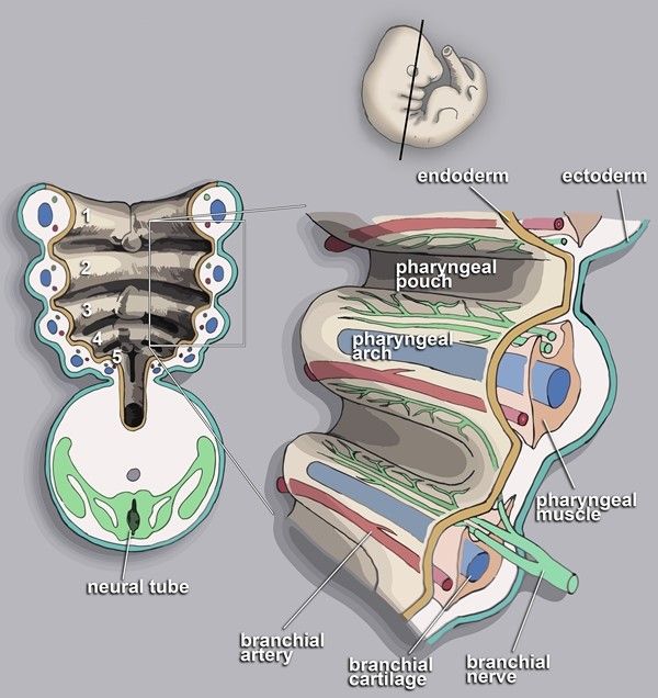
Each arch has a mesenchyme centre, an ectoderm outer covering, and an endoderm lining on the inside (next to the gut tube). Each arch also has an associated artery, nerve and cartilaginous bar that forms within it. The first arch has two discernible regions to it, named the maxillary and mandibular regions.
The recession between each arch on the inner side (the side lined with endoderm) is referred to as a pouch, and the recession between each arch on the outer side (the side lined with ectoderm) is referred to as a cleft.
Along with the pharyngeal arches, there is another structure named the frontonasal prominence; a single, unpaired structure that sits above the developing forebrain. Together, the frontonasal prominence (FNP) and five pairs of pharyngeal arches (PA) make up the developing elements in the head and neck region.

Diagram - The developing pharyngeal arches, along with their internal structure: each arch is associated with an artery, a nerve cartilage and muscle
Creative commons source by Loki austanfell, edited by Maddie Swannack [CC BY-SA 4.0 (https://creativecommons.org/licenses/by-sa/4.0)]
Nervous Derivative of the Pharyngeal Arches
There are number of cranial nerves that are associated with the pharyngeal arches – CN V, VII, IX and X.
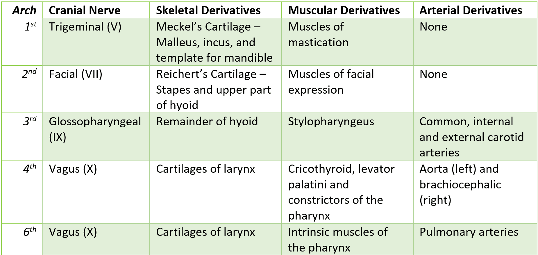
The trigeminal nerve (CN V) is associated with the first pharyngeal arch, and thus all derivatives of the first pharyngeal arch will be innervated by the trigeminal nerve. The same holds true for the second pharyngeal arch with the facial nerve (CN VII), the third pharyngeal arch with the glossopharyngeal nerve (CN IX), and the fourth and sixth pharyngeal arches with the vagus nerve (CN X).
Muscular Derivatives of the Pharyngeal Arches
As spoken about in the article ‘Muscles of the Head and Neck’ there are two groups of muscles in the face: the muscles of facial expression and the muscles of mastication.
The muscles of mastication are derived from the first pharyngeal arch (explaining why they are innervated by the mandibular branch of the trigeminal nerve) and the muscles of facial expression are derived from the second pharyngeal arch (fitting with their innervation from the facial nerve).
The third pharyngeal arch gives rise to the stylopharyngeus muscle, which acts to elevate the larynx and pharynx, and this is the only muscle in the pharynx innervated by the glossopharyngeal nerve.
The fourth pharyngeal arch is the origin of cricothyroid, levator palatini and the constrictors of the pharynx. Finally, the sixth arch is where the intrinsic muscles of the larynx originate. These are all innervated by the vagus nerve because this is the cranial nerve associated with the fourth and sixth pharyngeal arches.
Cartilage Derivatives of the Pharyngeal Arches
The cartilage bars in each pharyngeal arch give risk to the skeletal elements of the head and neck are derived from. Just like the pharyngeal arch, the pharyngeal cartilages decrease in size moving cranial to caudal. The first and second are large enough to get their own names.
Meckel’s cartilage comes from the first pharyngeal arch. This gives rise to the malleus and incus (two of the inner ear ossicles), as well as the template used for the mandible.
Reichert’s cartilage is the name of the second pharyngeal cartilage, supplying the stapes (third inner ear ossicle) along with the upper part of the hyoid bone.
The remainder of the hyoid bone is derived from the third pharyngeal cartilage. Pharyngeal arches four and six contribute to the cartilages of the larynx.
Arterial Derivatives of the Pharyngeal Arches
Before any major developmental processes take place, the arterial derivatives of the pharyngeal arches are essentially symmetrical and well structured. However, a number of shunts are required for the embryo to continue to develop, so remodelling must take place.
The arterial derivatives are a little easier to remember as the arterial components of the first two arches completely disappear.
The third becomes the internal carotid artery (on both sides of the midline).
The fourth pharyngeal arch forms the arch of the aorta on the left and the brachiocephalic artery on the right.
The sixth pharyngeal arch becomes the pulmonary arteries.
Summary of the Pharyngeal Arches

Table - A summary of the pharyngeal arches and their derivatives
As mentioned above, the pharyngeal pouches are the inner spaces between the pharyngeal arches that form pockets of endoderm in the pharynx.
As with the arches, the first pouch is the biggest, and ultimately becomes the tympanic cavity (the space where the inner ear ossicles are going to sit).
Moving caudally, the palatine tonsils (in combination with some lymphoid cells) originate from the second pharyngeal pouch. The parathyroid glands and the thymus (one lobe from either side fusing together) develop from the third and fourth pharyngeal pouches.
Normally, only the first cleft is what remains, as the external acoustic meatus. The remaining clefts are obliterated by the second cleft moving down over them.
On occasion, either a branchial cyst (enclosed fluid filled remnant) or fistula (abnormal opening from a branchial cyst to the neck) can occur from mis-obliteration of the clefts. These can present with various neck lumps, as explained in the article ‘Lymph Nodes and Neck Lumps’.
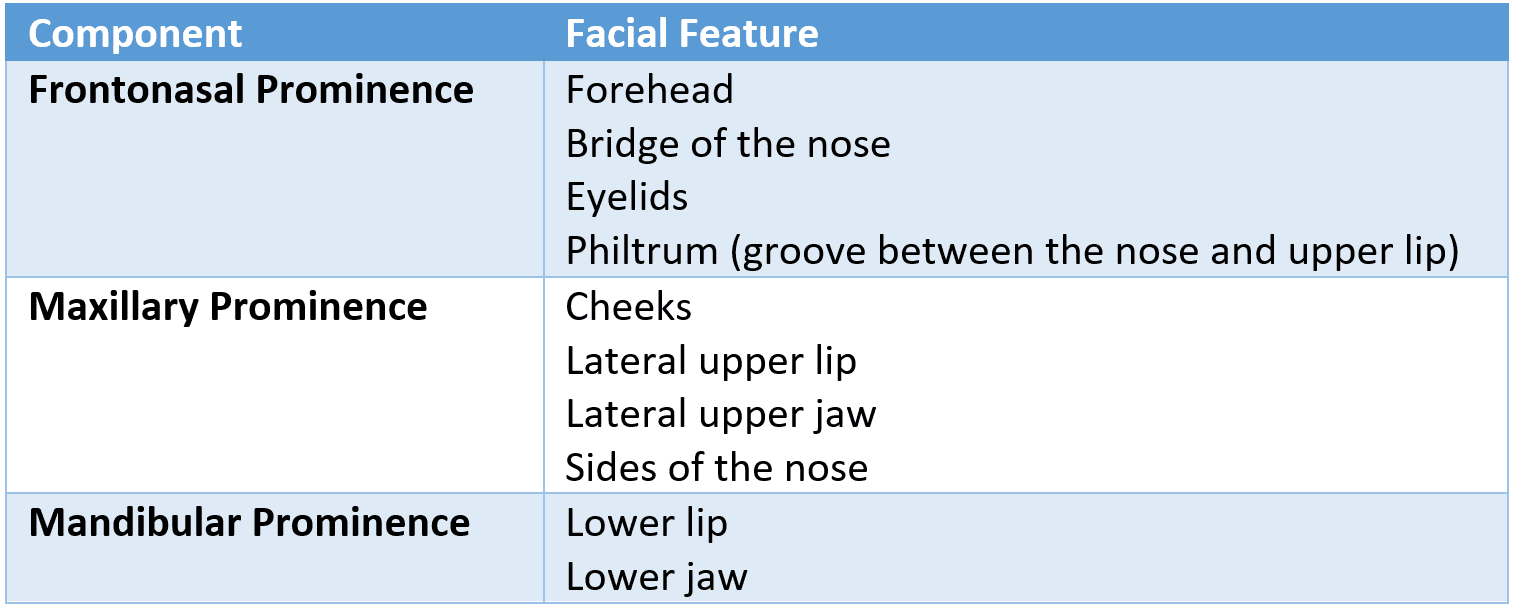
The key components for the development of the face are:
- The frontonasal prominence
- Two maxillary prominences (one from each side)
- Two mandibular prominences (one from each side)
The stomodeum also contributes to the development of the face. This is the end of the foregut that will form the mouth after the rupture of the buccopharyngeal membrane.
These components will then form the follow features:

Table - The facial features that develop from each facial prominence
Development of the Nose and Palate
Before discussing the development of the nose, the term placode needs to be introduced. Placodes are ectodermal thickenings that form in the cranial region of the embryo on either side of the frontonasal prominence. They will differentiate and develop to form the special sensory structures of the nose.
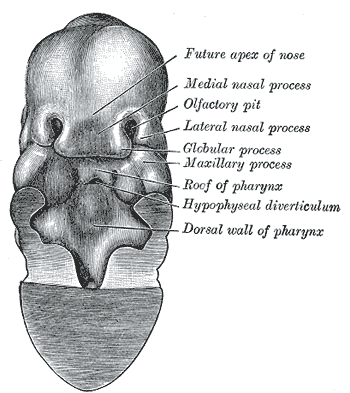
The nasal placodes then invaginate to form the olfactory pits, which will go on to become the nostrils. The olfactory pits lie dorsal to the stomodeum, separated by the oronasal membrane, which will eventually rupture so the oral and nasal cavities become one space.
During development, a horseshoe-shaped ridge forms around the olfactory pits, with the ‘arms’ of the horseshoe being the medial and lateral prominences. The medial prominences fuse together at the midline, creating the philtrum of the upper lip (the groove between the centre of the nose and the upper lip) and the primary palate (a small structure just internal to the medial prominences), and pushing the maxillary prominences laterally.
Internally, two palatal shelves grow from the internal side of the maxillary prominences. As they grow, they fuse at the midline with each other, and with the primary palate, forming the secondary palate and separating the oral and nasal cavities.

Diagram - The ventral (front) surface of the embryo with the developing face. The most important parts to note are the medial and lateral nasal processes, the olfactory pits and the maxillary process
Public Domain Source by Henry Gray: Anatomy of the Human Body (20th edition) [Public domain]
Like any other process in the human body, separation of the nasal and oral cavities can go wrong. Specifically:
- Lateral Cleft Lip – failure of fusion of the medial nasal prominence and maxillary prominence unilaterally (can also extend into the primary palate/intermaxillary segment).
- Cleft Lip and Palate – combined failure of palatal shelves to meet in the midline.
These defects are usually surgically repaired, and will need extensive follow up with speech and language therapists to aid with feeding and speech development.
Development of the Pituitary Gland
The pituitary is made up two different tissue types, the anterior and posterior pituitary glands.
The first is ectoderm, which grows as a protrusion upwards from the roof of the mouth. This is called Rathke’s pouch and will go on to develop into the anterior pituitary gland. As the skull develops, the roof of the mouth forms, and this pinches off the connection between the pituitary gland and the mouth.
The second is neuroectoderm, which grows as a protrusion from the diencephalon of the developing brain. This is called the infundibulum and will develop into the posterior pituitary gland.
The tissue type that each gland develops from reflects its function later in life, with the anterior lobe having an endocrine function as it developed from ectoderm, and the posterior lobe having a neuroendocrine function due to developing from neuroectoderm.
The precursor to the tongue appears on the floor of the oral cavity at roughly the same time as the palate begins to form (see above). Unlike most of what has been discussed above, the tongue does not derive from one pharyngeal arch, but is composed of elements from each arch to create a single, midline structure.
A number of changes occur on the floor of the oral cavity during the formation of the tongue.
Two lateral lingual swellings arise from tissue of the first pharyngeal arch, along with three median lingual swellings (the tuberculum impar, cupola and epiglottal swelling) which arise from a combination of the first to fourth arches.
During the fourth week of development, the lateral lingual swellings overgrow the tuberculum impar and they fuse together, with their line of fusion being the median sulcus of the tongue.
The contributions from the second and third arches fuse, with their line of fusion marked by a V-shaped groove, at the centre of which is the foramen caecum. This pit represents the origin of the thyroid gland.
The tongue forms at the floor of the oral cavity. With a program of apoptosis, it is freed from the floor, leaving the lingual frenulum as the only attachment. If this frenulum is too short, the resulting condition is called ankyloglossia, or ‘tongue tie’.
The innervation of the tongue is quite complex:
- Motor innervation is provided by the hypoglossal nerve (CN XII).
- General sensory innervation is provided by the lingual branch of the trigeminal nerve (CN V) in the anterior 2/3, and the glossopharyngeal nerve (CN IX) in the posterior 1/3.
- Special sensory innervation (taste) is provided by the chorda tympani branch of the facial nerve (CN VII) in the anterior 2/3, and the glossopharyngeal nerve (CN IX) in the posterior 1/3.
Development of the Thyroid Gland
The primordium of the thyroid gland begins on the floor of the oral cavity between the tuberculum impar and the cupola (later called the foramen cecum).
This developing thyroid tissue splits to form a bi-lobed diverticulum which then descends down through the embryonic neck. The lobes of the thyroid are connected by the isthmus.
During its descent, the thyroid is still connected to the tongue by the thyroglossal duct. Once the thyroid migrates, this duct should close. If it does not fully obliterate, thyroglossal duct cysts can form.
Reviewed by: Dr. Thomas Burnell
- 5970

