Next Lesson - Conditions of the Ankle
Core
Want more information on anatomy before you begin looking at conditions? Head to our article on Bones of the Foot for more information!
Mallet Toe is flexion at the distal interphalangeal joint (DIPJ), and Hammer Toe is flexion at the proximal interphalangeal joint (PIPJ). Both of these conditions are most common in the second toe. They are caused by ill-fitting shoes or pressure from the adjacent first toe having Hallux Valgus (lateral deviation of the Hallux). The change in shape of the toe can cause shoes to rub against it and this can lead to painful corns or calluses.
Management options include using inserts or pads to reposition the toe when wearing footwear (this will help to relive pressure and pain - this works best if the toe is still flexible); if this more conservative treatment is unsuccessful, surgery may be recommended to straighten/flatten the toe.
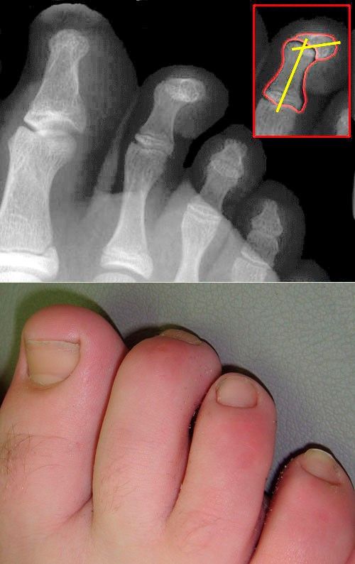
Image - X-ray and Physical Appearance of Mallet Toe
Creative commons source by J. Lengerke [CC BY-SA 3.0 de (https://creativecommons.org/licenses/by-sa/3.0/de/deed.en)]
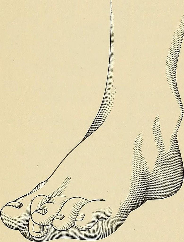
Image - Drawing of Hammer Toe
Creative commons source by FREDERIC FRANCIS BURGHARD, Operative Surgery (1909), Page 599 [CC BY-SA 3.0 de (https://creativecommons.org/licenses/by-sa/3.0/de/deed.en)]
Claw Toe describes hyperextension of the metatarsal phalangeal joint (MTPJ) and flexion of the proximal interphalangeal joint (PIPJ) and sometimes also flexion at the distal interphalangeal joint (DIPJ). It often affects all 4 lateral toes at the same time and usually occurs secondarily to neurological damage, trauma or Rheumatoid Arthritis.
The condition is split into two categories:
- Flexible Claw Toe – where the joints remain mobile and pain is less severe.
- Rigid Claw Toe – a fixed deformity that is very painful.
Flexible Claw Toe is treated by taping the affected toes/wearing a splint to keep the toes in the right position and performing exercises to maintain toe flexibility. Rigid Claw Toe is treated with surgery, where the bone is shortened at the base of the toe to give them more room to straighten out.
Hallux Valgus is the medial deviation of the first metatarsal, lateral deviation of the hallux and prominence of the first metatarsal head. It is commonly referred to as a ‘Bunion’. This condition is most common in middle aged females and is caused by abnormal mechanics of over-pronated feet (meaning more weight is transmitted through the medial side of the foot than the lateral side). This is the most common foot deformity.
Management for this condition involves wearing wide shoes with low heels and soft soles, losing weight if overweight, and managing pain with over the counter medication. If the pain caused by Hallux Valgus is very severe and chronic, surgery can be used to remove bunions (commonly by removing the head of the metatarsal), however, this only occurs if the condition is having a large effect on the patients’ life. This surgery is not done cosmetically on the NHS.
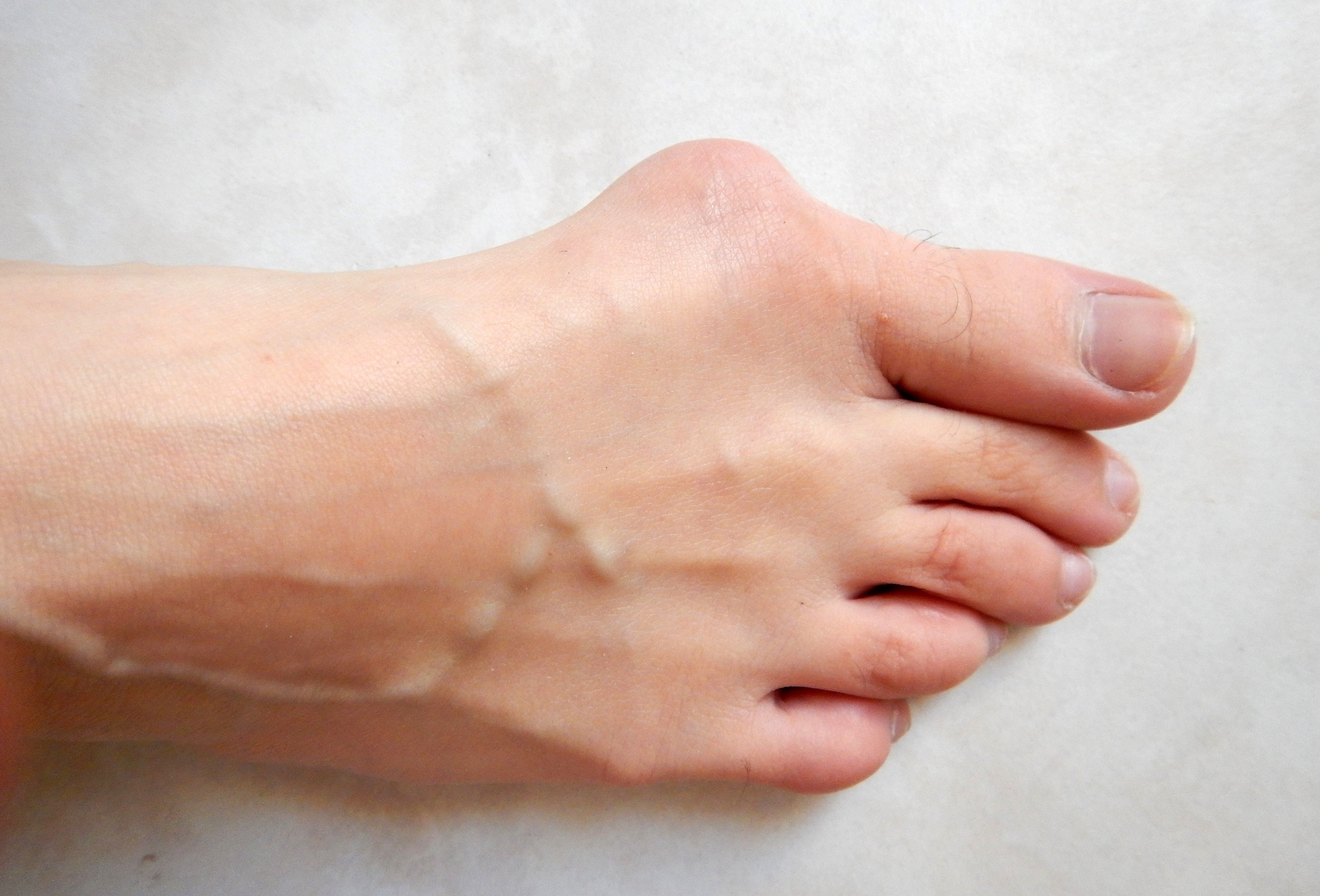
Image - Hallux Valgus
Creative commons source by Lamiot [CC BY-SA 4.0 (https://creativecommons.org/licenses/by-sa/4.0)]
Hallux Rigidus is osteoarthritis of the metatarsal joint of the hallux. This joint is very prone to osteoarthritis, due to the forces are transmitted through the foot on walking, so this is a common condition. The patient usually presents with pain in the metatarsal phalangeal joint (MTPJ) upon flexion of the hallux. In severe Hallux Rigidus, X-rays can be used to show the extent of the osteoarthritis order to decide if surgical intervention is required. However, normal management of the condition is to alter the patient's shoes to take the strain off the hallux when walking.
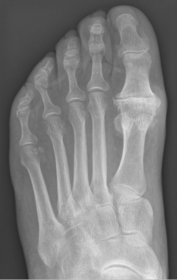
Image - X-ray Appearance of Severe Hallux Rigidus
Creative commons source by Hellerhoff [CC BY-SA 3.0 (https://creativecommons.org/licenses/by-sa/3.0)]
Pes Planus, also known as flat foot, is the collapse of the medial arch of the foot. It is important to note this condition is physiologically normal in children as the arch is still developing. In adults, it is caused due to stretching of the plantar calcaneonavicular ligament and plantar aponeurosis. This condition does not require treatment unless it is causing pain or restricting movement enough to affect daily life. If this is the case, exercises and specialised shoes may be recommended (surgical intervention is incredibly rare).
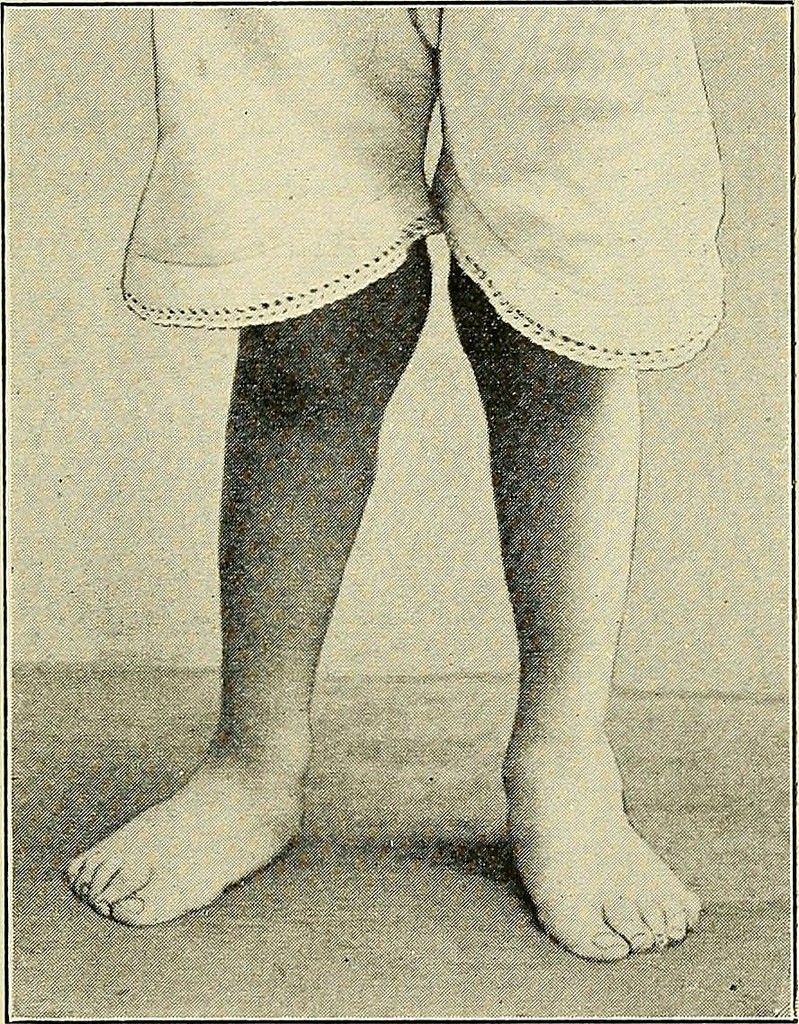
Image - Photograph of Pes Planus
Creative commons source by WHITMAN, ROYAL, A Treatise on Orthopedic Surgery (1910), Page 720 [CC BY-SA 3.0 de (https://creativecommons.org/licenses/by-sa/3.0/de/deed.en)]
Charcot Arthropathy is the progressive destruction of bones, joints and soft tissues secondary to Diabetes Mellitus. This most commonly occurs in the feet and ankle but is generally considered a rare condition. Neuropathy causes reduced sensation in the feet, which means any damage/injury to the feet may worsen without the patient being aware. Neuropathy can also lead to muscle spasticity, causing deformities. When the midfoot is involved in Charcot Arthropathy, Rocker-bottom foot (prominent calcaneus and convex/rounded bottom of the foot) can develop. Treatment can either be staying off the foot for 2-3 months or surgery.
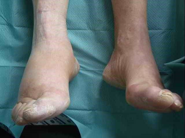
Image - A Patient with Charcot Arthropathy
Creative commons source by Radschläger13 [CC BY-SA 4.0 (https://creativecommons.org/licenses/by-sa/4.0)]
Edited by: Dr. Maddie Swannack
Reviewed by: Dr. Marcus Judge
- 6535

