Next Lesson - Bones of the Upper Limb
Abstract
The leg is split into three muscle compartments:
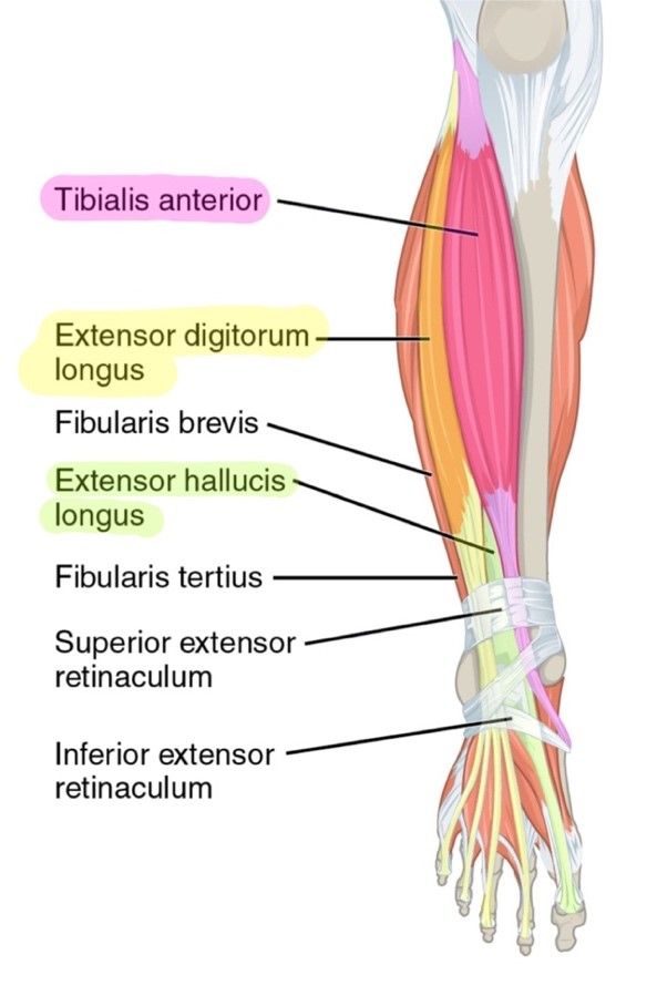
- Anterior compartment – dorsiflexes the foot and is innervated by the deep fibular nerve. The compartment contains tibialis anterior, extensor digitorum longus and extensor hallucis longus.
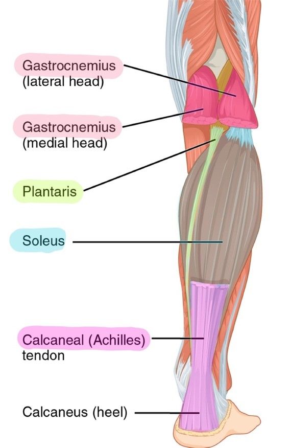
- Posterior compartment – plantarflexes the foot and is innervated by the tibial nerve. The compartment contains gastrocnemius, soleus, plantaris, popliteus, tibialis posterior, flexor digitorum longus and flexor hallucis longus.
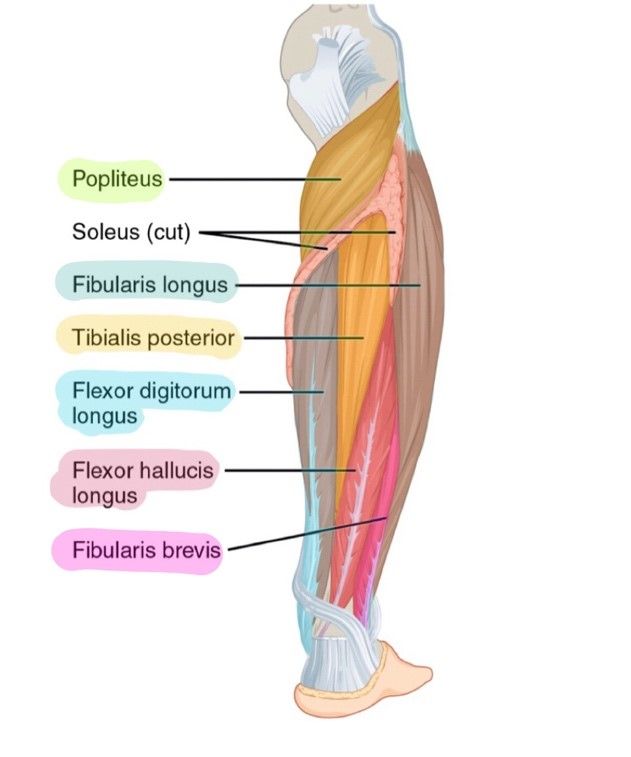
- Lateral compartment – everts the foot and is innervated by the superficial fibular nerve. The compartment contains fibularis longus and fibularis brevis.
Core
Muscles of the lower leg typically act at the ankle joint and are innervated by branches of the sciatic nerve. Whilst there are many muscles in the leg, they can be compartmentalised into anterior, lateral and posterior fascial compartments.
It is also very important when studying muscles that you are comfortable with anatomical terminology, as a lot is used in these articles. For more information, please check out our Anatomical Terminology Article.
Muscles in the anterior compartment act to dorsiflex the foot and are innervated by the deep fibular nerve (a branch of the common fibular nerve).
Tibialis Anterior is found on the lateral surface of the tibia, where it originates. It attaches to the medial cuneiform (one of the bones in the foot) and acts to dorsiflex the foot. It also acts to invert the foot.
Extensor Digitorum Longus originates from the lateral condyle of the tibia and forms four tendons, which insert into a digit (with exception of the hallux/big toe). As the name suggests, these act to extend the toes, whilst also aiding dorsiflexion.
Extensor Hallucis Longus originates from the medial fibula and inserts into the distal phalanx of the big toe. It is responsible for the extension of the hallux/big toe but also contributes to dorsiflexion of the foot.

Diagram - Anterior compartment of muscles in the leg, highlighted. Notice how deep the extensor hallucis longus is compared to the tibialis anterior and extensor digitorum longus
Creative commons source by OpenStax College, edited by Sachin Sudhakaran [CC BY-SA 4.0 (https://creativecommons.org/licenses/by-sa/4.0)]
The posterior compartment is slightly more complex, in that it is split into superficial and deep layers. Both layers are innervated by the tibial nerve and typically act to plantarflex the foot.
Superficial Muscles of the Posterior Compartment
Each superficial muscle inserts into the calcaneus of the foot via the calcaneal tendon (aka. Achilles tendon). Each of these muscles are innervated by the tibial nerve. There are three main muscles to know:
Gastrocnemius is the most superficial of the posterior leg muscles. It has a medial and lateral head, which originate from the medial and lateral femoral condyles respectively. These converge to form single muscle belly, which then attaches to the calcaneus as described above. It acts to plantarflex the foot. It can also aid knee flexion as it crosses the knee joint.
A large, flat muscles located just deep to Gastrocnemius, Soleus gets it name from being shaped like a sole (a big flat fish). It originates from the soleal line of the tibia and attaches to the calcaneus. It acts to plantarflex the foot.
Found just superficial to the soleus, it is a small muscle with a very long tendon. It originates from the lateral epicondyle of the femur and attaches via the calcaneal tendon. Like the other muscles, it acts to plantarflex the foot.
Deep Muscles of the Posterior Compartment
The remaining muscles of the posterior compartment are deep. Notice the similarities with the anterior compartment of leg muscles. In the deep posterior compartment, there is a tibialis muscle which acts to plantarflex and invert the foot, there’s a muscle for the flexion of the toes and a muscle for flexion of the big toe. With exception of the popliteus, it’s useful to know that tendons of the muscles in the deep posterior compartment travel posterior to the medial malleolus, allowing access to the inferior surface of the foot for attachment. Once again, each muscle is innervated by the tibial nerve. There are four main muscles to know:
This muscle lies posterior to the knee joint. It originates from the lateral femoral condyle and attaches just above the soleal line of the tibia. It is unique in that it acts to 'unlock the knee' by laterally rotate the femur on the tibia; normally when standing, this locking allows for the knee to support the standing weight of a human, and so popliteus is important for unlocking the knee so knee flexion can occur.
A large, deep muscle which originates between the tibia and fibula, via the interosseous membrane. It attaches to the inferior surface of the medial tarsal bones. This acts to plantarflex and invert the foot.
This muscle is located medially in the posterior compartment. It originates from the medial tibia and attaches to the inferior surface of each of the four digits. It acts to flex each toe (with exception of the hallux).
This muscle is located laterally in the posterior compartment, and originates from the fibula. It attaches to the inferior surface of the distal phalanx of the hallux. As the name suggests, it acts to flex the hallux.

Diagram - Superficial posterior compartment of the leg, highlighted. Notice how long the tendon of plantaris is
Creative commons source by OpenStax College, edited by Sachin Sudhakaran [CC BY-SA 4.0 (https://creativecommons.org/licenses/by-sa/4.0)]
The muscles in the lateral compartment of the leg are responsible for everting the foot. The muscles are innervated by the superficial fibular nerve (a branch of the common fibular nerve) and the tendons pass posterior to the lateral malleolus.
A long muscle that originates from the lateral surface of the fibula, the Fibularis Longus descends posterior to the lateral malleolus and attaches to the medial cuneiform. It everts the foot, whilst also aiding plantarflexion.
Fibularis Brevis is a small muscle that is found inferior and deep to the fibularis longus muscle. It arises from the lateral fibula and attaches to the fifth metatarsal. It solely acts to evert the foot.

Diagram - Deep posterior and lateral compartment of the leg, highlighted
Creative commons source by OpenStax College, edited by Sachin Sudhakaran [CC BY-SA 4.0 (https://creativecommons.org/licenses/by-sa/4.0)]
For Pathologies and Conditions affecting the lower leg area, check out our Conditions of the Lower Leg article.
Reviewed by: Dr. Thomas Burnell
Edited by: Dr. Maddie Swannack
- 28140

