By Emily Smith
Next Lesson - Anatomy of the Woman
Core
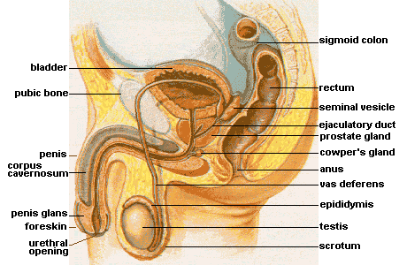
Diagram - Anatomy of the male reproductive system
Creative commons source by alt.sex FAQ, © Elf Sternberg [CC BY-SA 4.0 (https://creativecommons.org/licenses/by-sa/4.0)]
The function of the penis includes:
- Expulsion of urine through the urethra
- Deposition of sperm
The penis is made up of two key erectile tissues.
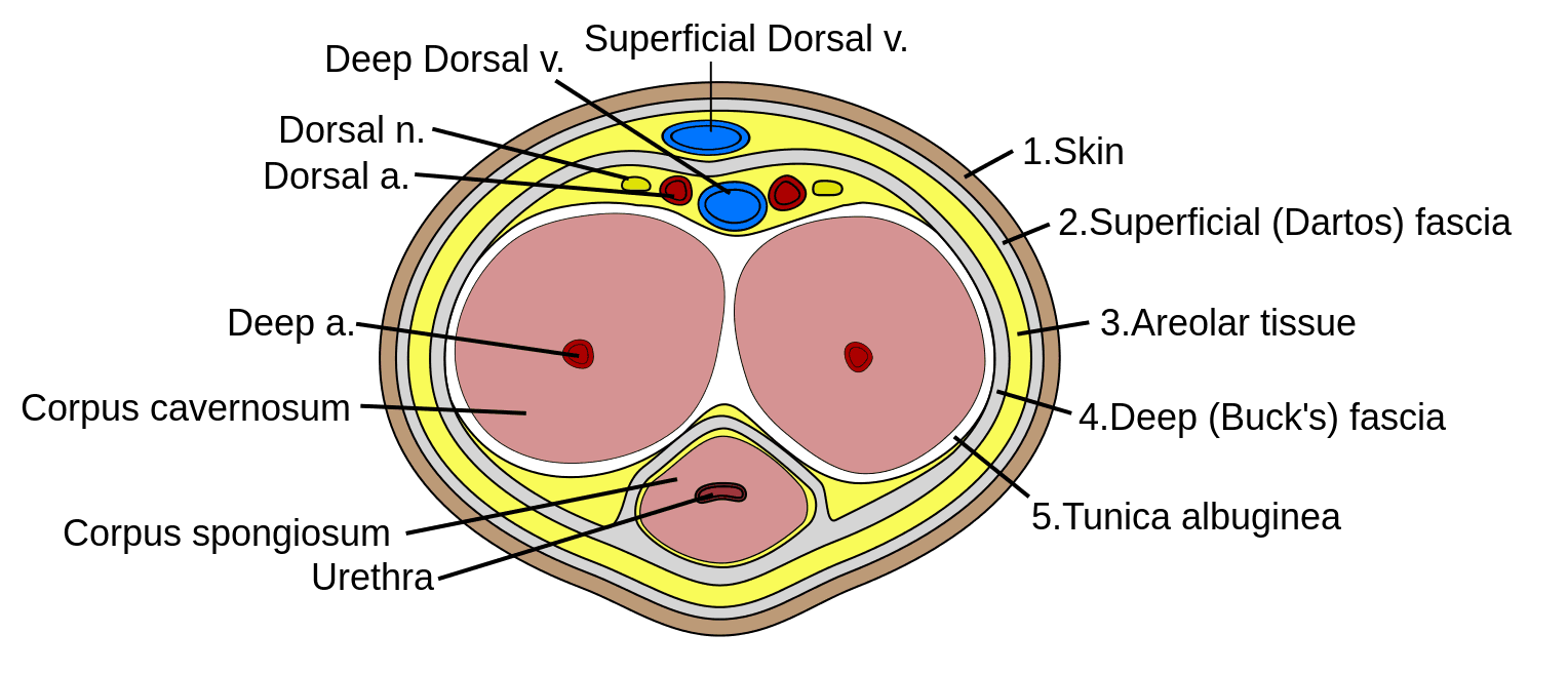
Diagram - Cross section of the penis showing the erectile tissue
Creative commons source by Mcstrother [CC BY-SA 4.0 (https://creativecommons.org/licenses/by-sa/4.0)]
The corpus cavernosum is the main erectile tissue of the penis. There are two columns of this tissue type in the penis, and they are anchored at the pubic symphysis. The corpus spongiosum is an expansible tissue which communicates the urethra, it is of lesser importance in producing an erection.
An erection is stimulated by the parasympathetic nervous system. It causes vasodilation of arterioles and relaxation of the sinusoids of the corpus cavernosa.
Ejaculation is stimulated by the sympathetic nervous system. This involves contraction of glands and ducts of the male reproductive system to expel sperm, and the closing of the internal urethral sphincter to prevent retrograde ejaculation into the bladder.
This can be remembered by the rather crude ‘Point and Shoot’ which indicates that erection is achieved by the parasympathetic system (P for point) and that ejaculation is achieved by the sympathetic system (S for shoot).
Erectile dysfunction is the inability to attain and maintain an erection. Its causes can be classified as organic, psychosexual, or mixed. The most common cause is vascular due to reduced blood flow due to cardiovascular disease, hypertension, diabetes mellitus, or smoking. Erectile dysfunction can also be a side effect of some medications.
The tunica albuginea surrounds each of the corpora cavernosa and gives the penis its cylindrical shape on erection. Damage to this tissue is known as a penile fracture and leads to deformation of the penis on erection.
The clitoris is the embryological equivalent to the penis in females.
Blood supply to the penis is from the internal pudendal artery which is a branch of the internal iliac artery.
In the embryo, the testes begin their development at the posterior abdominal wall and descend to their position in the scrotum. This allows spermatogenesis to occur at the optimum temperature which is just lower than body temperature.
The descent of the testes creates the inguinal canal as it brings down the layers of the abdominal wall. They also bring down the peritoneum which surrounds the testes forming the tunica vaginalis. This has a parietal and visceral layer as it is formed from the peritoneum and allows for friction-free movement of the testes; these can fill with excess fluid forming a hydrocoele.
The tunica albuginea is a fibrous capsule surrounding the testes which divides the parenchyma into lobules.
The seminiferous tubules are lined with Sertoli cells; these are the site of spermatogenesis. The sperm travels along here to the rete testis, then to the epididymis – this is the site in which the maturation of sperm occurs. The epididymis then goes on to form the vas deferens (or ductus deferens) which is the component that travels through the spermatic cord.
The Leydig cells of the testicle surround the Sertoli cells, and they produce testosterone.
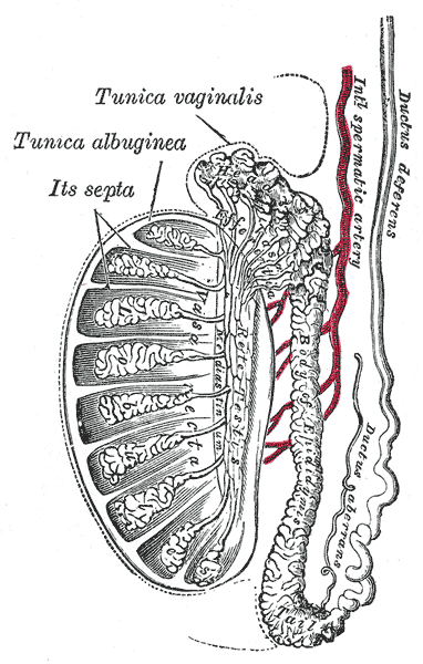
Diagram - The anatomy of the testis
Public Domain Source by Henry Gray (1918) Anatomy of the Human Body [Public domain]
The testes receive their blood supply from the testicular arteries, which arise directly from the abdominal aorta. These arteries are long due to the descent of the testes.
The venous drainage is by the pampiniform plexus which then forms the testicular vein. On the right, the blood drains directly from the pampiniform plexus into the inferior vena cava, whereas the left drains into the left renal vein. These veins also surround the testicular arteries; this allows the transfer of heat to cool down blood entering the testes to allow for the cooler temperature required for spermatogenesis.
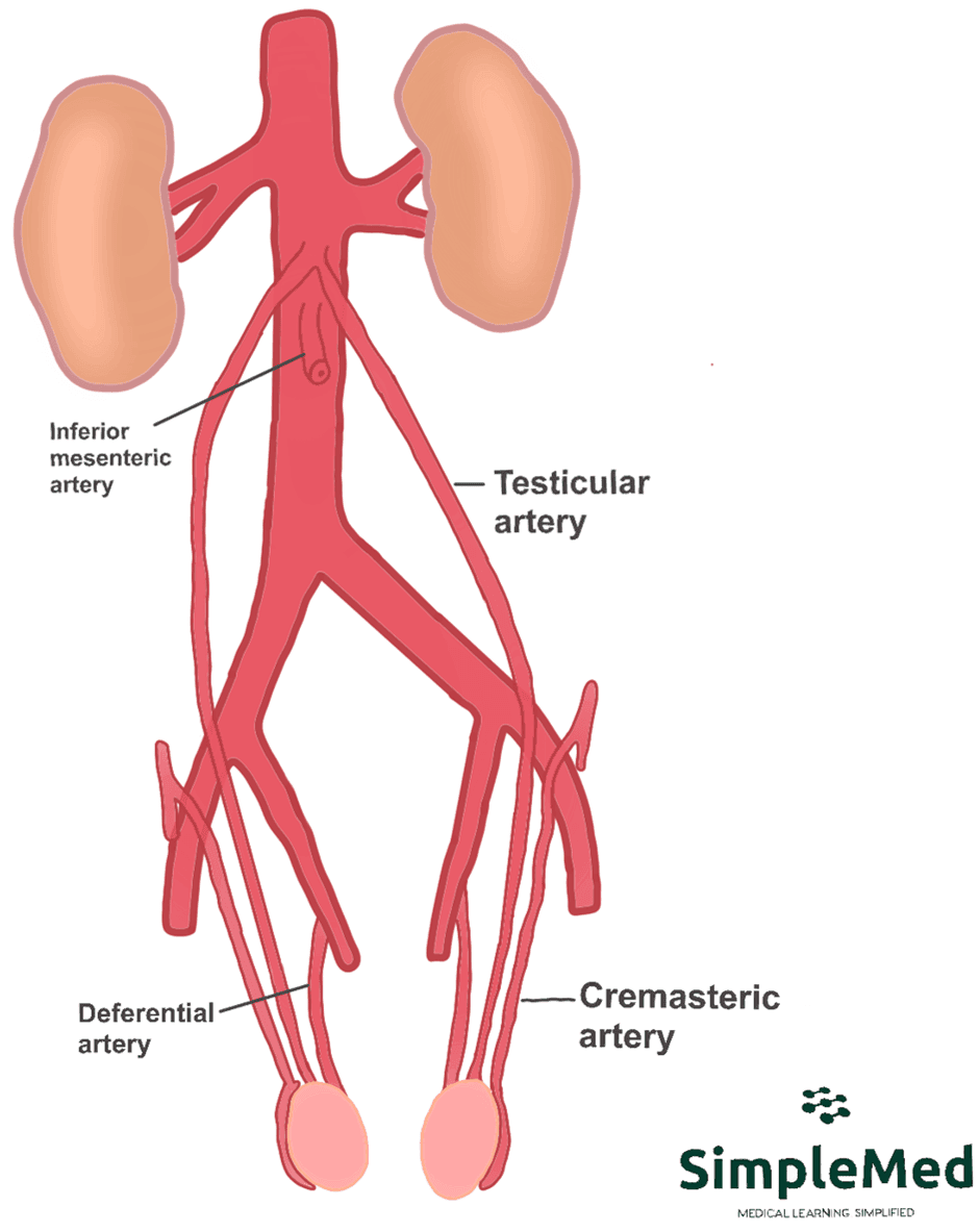
Diagram - The arterial supply to the testes
SimpleMed original by Emily Smith
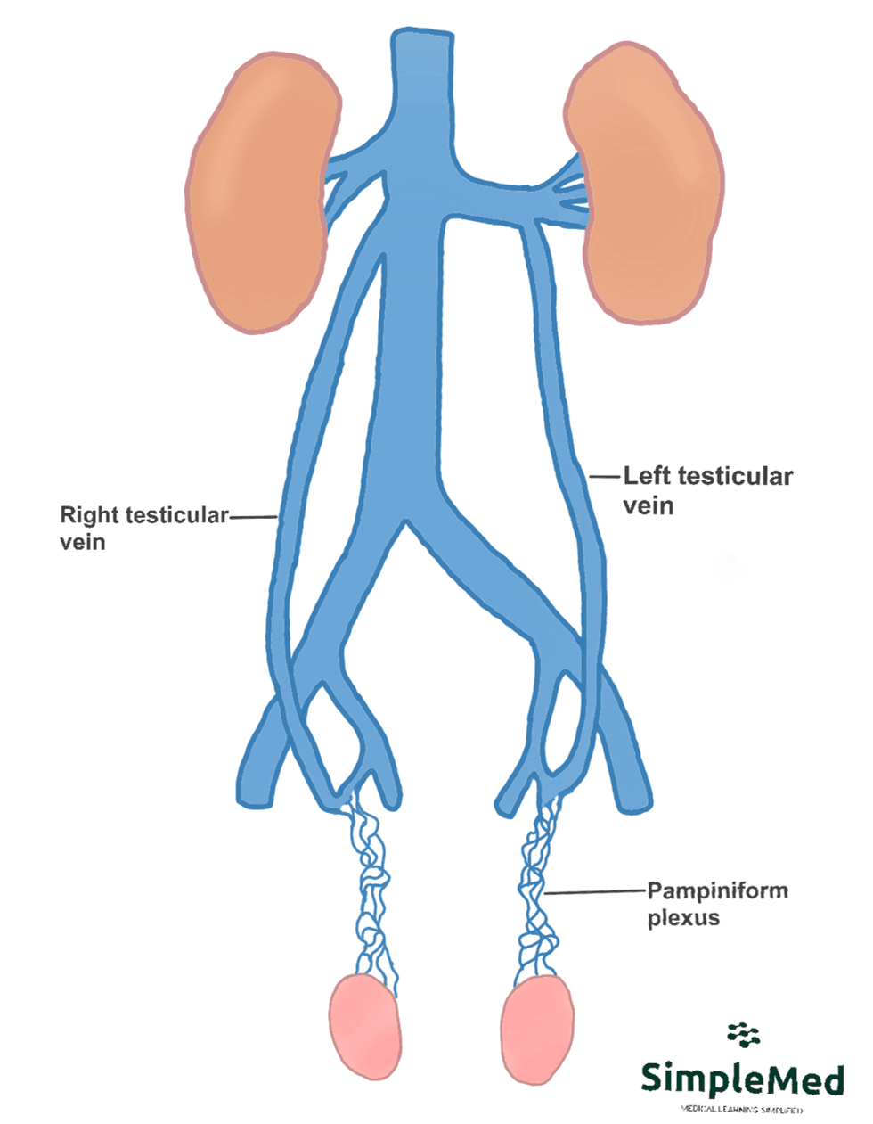
Diagram - The venous drainage of the testicles. Note that the left pampiniform plexus drains into the left renal vein, and the right pampiniform plexus drains straight into the inferior vena cava
SimpleMed original by Emily Smith
Obstruction of the testicular veins can lead to a varicocele. This is swelling of the scrotum due to increased hydrostatic pressure within the pampiniform plexus. Primary causes of this include incompetent or absent valves. They can also occur due to renal pathology or tumours compressing the veins. They most commonly occur on the left side due to the route taken by the veins. They are typically painless and the diagnosis is clinical, although an ultrasound scan can also be carried out to help with diagnosis. Minor cases don’t require treatment whereas more serious cases may require surgery.
The spermatic cord provides the neurovascular supply to the testes. It begins its course at the deep inguinal ring, then passes through the inguinal canal and enters the scrotum through the superficial inguinal ring.
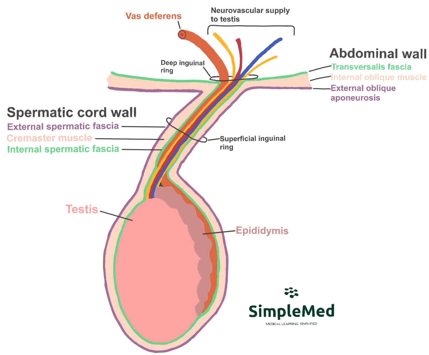
Diagram - The structure of the spermatic cord
SimpleMed original by Emily Smith
The contents of the spermatic cord includes:
- 3 Fascial Layers
- External spermatic fascia
- Cremaster muscle and fascia
- Internal spermatic fascia
- 3 Arteries
- Testicular artery
- Artery to vas deferens
- Cremasteric artery
- 3 Veins
- Pampiniform plexus
- Vein to vas deferens
- Cremasteric vein
- 3 Nerves
- Genital branch of genitofemoral nerve
- Sympathetic nerve to vas deferens
- Cremasteric nerve
- Vas Deferens
- Lymphatics – these are the para-aortic lymph nodes which drain the testes.
- Processus Vaginalis – this is a remnant of the peritoneum from the descent of the testes in embryonic development.
The spermatic cord can become twisted on itself resulting in testicular torsion. This requires surgery as soon as possible as there is a risk of necrosis of the testis. A risk factor for this is a ‘bell clapper deformity’ which is where the testis is able to move within the tunica vaginalis. Patients with this present with sudden onset, severe pain in one testis. They often also have nausea and vomiting. Easing of this pain can indicate the start of necrosis.
These glands produce the majority of the ejaculate – approximately 65%. The fluid produced is alkaline to neutralise the acidic environment of the vagina, and provides nutrition to sperm.
This gland produces about 25% of the ejaculate. This secretion contains proteolytic enzymes and is mildly acidic.
The prostate wraps around the urethra, so enlargement such as in benign prostatic hyperplasia can lead to urinary symptoms such as frequency, urgency, and incontinence. The prostate also sits just anterior to the rectum, meaning pathology of the prostate gland (e.g. prostate cancer) can be identified in a rectal exam (PR exam).
These glands only produce a small amount of the ejaculate which reduces friction.
Edited by: Dr. Ben Appleby and Dr. Maddie Swannack
Reviewed by: Dr. Thomas Burnell
- 7864

