Next Lesson - Gametogenesis
Contents
- The Hypothalamic-Pituitary-Ovarian Axis
- Menstrual Cycle Phases
- Early Ovarian Cycle
- Ovulation
- Uterine Cycle
- Amenorrhoea
- Menorrhagia
- Irregular Menstrual Bleeding
- Intermenstrual or Post-Coital Bleeding
- Dysmenorrhoea
- Premenstrual Syndrome
- Investigation and Management of Menstrual Disorders
- Quiz
- Feedback
Abstract
- The hypothalamic-pituitary-ovarian axis is the hormonal axis responsible for controlling the menstrual cycle.
- Differing levels of hormones in the HPO axis are responsible for the initiation of ovulation and the shedding of the endometrium at the end of the cycle.
- Problems with menstruation can present with amenorrhoea, oligomenorrhoea, or menorrhagia.
- A patient-centred approach to the investigation and management of menstrual disorders is key to achieving good outcomes for patients.
Core
The Hypothalamic-Pituitary-Ovarian Axis
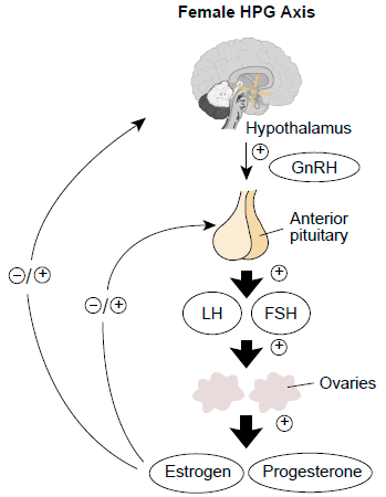
The menstrual cycle is regulated by the hypothalamic-pituitary-ovarian (HPO) axis. The hypothalamus releases gonadotrophin-releasing hormone (GnRH) which stimulates the anterior pituitary gland to release the gonadotrophins luteinising hormone (LH) and follicle-stimulating hormone (FSH), which in turn stimulates the ovaries to produce oestrogen, progesterone and inhibin.
Oestrogen and progesterone can either positively or negatively feedback on the anterior pituitary and hypothalamus in order to control the axis. Inhibin release is prompted by the release of FSH, and acts to inhibit further release of FSH to prevent more than one follicle being stimulated for release at once.
GnRH is released in a pulsatile way (meaning it is not continuously released). This is because GnRH receptors which are continuously exposed to GnRH become desensitised which causes LH, FSH, oestrogen, and progesterone production to stop.

Diagram - The Hypothalamic-Pituitary-Ovarian axis
Creative commons source by Lu Kong, Ting Zhang, Meng Tang and Dayong Wang
[CC BY-SA 4.0 (https://creativecommons.org/licenses/by-sa/4.0)]
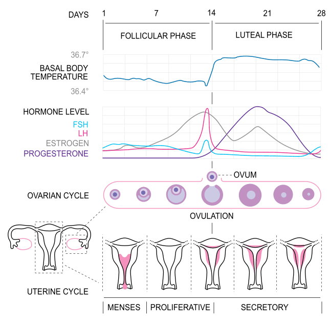
The menstrual cycle can be described by two smaller cycles by looking at two parts of the reproductive tract, the ovaries and the endometrium. The menstrual cycle is classically described as lasting 28 days, but this can vary from person to person.
The ovarian cycle is split into two phases: the follicular and luteal phases. Each phase in the ovarian cycle lasts 14 days.
The follicular phase begins at day 0 and involves the development of the ovarian follicle (containing the ovum). On day 14 the follicle ruptures and the ovum is released from the ovary, where it passes down the fallopian tube and into the uterus.
Over the next 14 days, the luteal phase occurs, and the follicle develops into the corpus luteum which will help to maintain an early pregnancy if fertilisation occurs. If fertilisation does not occur, the corpus luteum atrophies and becomes the non-functioning corpus albicans.
The endometrial cycle has three phases: menses, the proliferative phase, and the secretory phase.
Menses is the bleeding which begins on day 1 and represents the shedding of the endometrium in the absence of implantation taking place, this continues until around day 4 of the menstrual cycle.
The proliferative phase occurs from day 4 to day 14 and is where the endometrium lining is thickened in preparation for implantation under the influence of oestrogen.
Day 14-28 is when the secretory phase occurs; this is where the endometrium lining is at its thickest and contains glandular epithelium. This occurs under the influence of progesterone.

Diagram - A graph detailing the changes in hormones, endometrial changes, and ovarian changes throughout the menstrual cycle. Note: Inhibin levels not shown on the graph
Creative commons source by Isometrik [CC BY-SA 4.0 (https://creativecommons.org/licenses/by-sa/4.0)]
In the first few days of the menstrual cycle the levels of ovarian hormones are very low with a low level of inhibin. This means there is very little inhibition of the hypothalamus and anterior pituitary which causes FSH levels to rise. FSH binds to the granulosa cells of the developing ovarian follicle and stimulates it to continue development until the theca interna develops, which marks when the follicle is capable of producing oestrogen and inhibin.
In the mid-follicular phase, the follicle produces large amounts of oestrogen, which exerts a positive feedback effect on the hypothalamus and anterior pituitary; this causes an increase in LH and FSH. This occurs because in the presence of a high oestrogen concentration, LH levels are more sensitive to a pulse of GnRH meaning more LH is produced per GnRH pulse.
Before ovulation, oestrogen and inhibin rise rapidly as the follicle is developed enough to produce oestrogen without stimulation through FSH. This leads to a surge in LH production. The surge of LH causes the granulosa cells of the follicle to produce progesterone. Prompted by the LH surge, ovulation occurs, where the mature oocyte ruptures the capsule of the ovary and is released into the fallopian tube.
After ovulation, the follicle becomes ‘luteinised’ and continues to secrete large amounts of oestrogen and progesterone, and a small amount of inhibin. LH production is now suppressed as progesterone from the corpus luteum exerts a negative feedback effect. This causes further maturation of oocytes to be put on hold.
Without fertilisation, there is no further rise in LH, meaning the corpus luteum will regress spontaneously. This leads to a large fall of oestrogen and progesterone meaning the negative feedback on the HPO axis is removed and LH is free to rise again, and the cycle starts over.
If fertilisation occurs, the syncytiotrophoblast in the developing embryo produces human chorionic gonadotrophin (HCG) which acts to sustain the corpus luteum. This allows the corpus luteum to continue to produce oestrogen and progesterone which supports the pregnancy. Eventually the placenta becomes developed to the level at which it can produce enough oestrogen and progesterone to control the HPO axis throughout the pregnancy. These high levels of oestrogen and progesterone result in negative feedback on the HPO axis, preventing further maturation and release of oocytes.
The endometrium is sensitive to the ovarian hormones: oestrogen and progesterone. Oestrogen causes the proliferation of the endometrium, and oestrogen and progesterone cause the secretion of the endometrium.
The endometrium is split into two layers:
- Functional layer – the layer that proliferates and sheds at the end of the cycle.
- Basal layer – the layer where the cells in the functional layer grow from.
At the start of the proliferative phase, there are few glands in the functional layer of the endometrium. Throughout the proliferative phase, glands begin to coil and form in the thickened endometrium ready for implantation if fertilisation occurs. By the end of the proliferative phase, the functional layer has doubled in size. The secretory phase begins with the endometrium at its maximum thickness with very pronounced coiled glands. In the late secretory phase, the glands have a characteristic saw-tooth appearance.
The next phase of the uterine cycle is the menses. This is the phase during which the functional layer sheds, forming a period, leaving the basal layer behind to proliferate in the next cycle.
Amenorrhoea is the absence of menstruation.
Causes can be split into primary or secondary amenorrhea.
- Primary amenorrhoea is defined as the failure to establish menarche by 16 years of age.
- Secondary amenorrhoea is defined as the cessation of previously normal menstruation for a period of 6 months or more.
There are physiological causes for amenorrhoea such as being pre-pubertal, pregnancy, or post-menopause.
Pathological causes of amenorrhoea can occur at any level of the HPO axis (the hypothalamus, pituitary gland, or ovaries or with outflow (the uterus, cervix, or vagina).
The exact location of the pathology can be determined by looking at the levels of LH and FSH in the blood – low LH and FSH would suggest an issue with the hypothalamus or pituitary gland, whereas a normal/high LH and FSH would suggest an issue with the ovaries or outflow tract.
There are also structural causes of amenorrhoea where there is a mechanical obstruction of blood flow:
- Imperforate Hymen – a condition occurring when a thin layer of connective tissue (hymen) covers the vaginal entrance, and this has not perforated at the onset of menstruation. This can lead to a collection of blood behind the hymen, and progress to an infection.
- Cervical Stenosis – where the os of the cervix is not open meaning blood cannot flow from the uterus into the vaginal canal.
- Vaginal Septae – where a wall of tissue forms during the development of the vagina, separating the upper vagina into two parts, one of which forms a blind-ended pouch where blood can collect.
- Agenesis or Hypoplasia of the Genital Tract – underdevelopment (hypoplasia) or lack of development (agenesis) of any part of the genital tract can stop the movement of blood.
- Asherman’s Syndrome – an acquired condition where scar tissue forms in the uterus, often following instrumentation of the uterus such as IUS insertion or surgical termination, blocking the flow of blood.
- Uterine Fibroids – large uterine fibroids (leiomyomas) can cause the blockage of blood in the uterus.
Menorrhagia or heavy menstrual bleeding (HMB) is the complaint of excessive menstrual blood loss over consecutive cycles or >80ml per menstruation.
Menorrhagia is a common presenting complaint with a wide range of differentials varying from trivial to very serious:
- Bleeding tendency, such as von Willebrand Disease
- Hypothyroidism
- Iatrogenic, such as due to anticoagulation or insertion of a copper intrauterine device (IUD).
- Uterine polyps
- Uterine fibroids
- Endometrial cancer
Irregular menstrual bleeding is another common gynaecological presentation.
Oligomenorrhoea is defined as infrequent menstruation with cycles lasting more than 35 days (having 4-9 periods per year). It can present as a change in a patient’s usual bleeding pattern and is very common with progesterone-only hormonal contraception.
Other causes include:
- Polycystic ovarian syndrome (PCOS) – a condition of irregular ovulation leading to irregular periods.
- PID
- Hyperthyroidism
- High levels of prolactin – this can have many causes including breastfeeding or the presence of a prolactin-secreting tumour.
Intermenstrual or Post-Coital Bleeding
Intermenstrual bleeding (IMB) is bleeding unrelated to a period that occurs within a regular cycle.
Post-coital bleeding (PCB) is bleeding unrelated to a period that occurs after sex.
Causes of IMB and PCB range from benign to easily treatable to malignant, so it is important to investigate thoroughly.
In a woman who has gone through the menopause and is no longer menstruating regularly, any sort of vaginal bleeding should be investigated for the causes below.
Causes include:
- Sexually Transmitted Infections (STIs)/Pelvic Inflammatory Disease (PID)
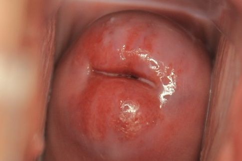
- Cervical ectropion – the evolution of the cells of the internal cervical canal to the outer surface of the cervix which can cause them to bleed.
- Cervical cancer
- Endometrial polyps or cancer
- Ovarian cysts

Image - A cervical ectropion as seen on speculum examination
Creative commons source by Gynpath.ru [CC BY-SA 4.0 (https://creativecommons.org/licenses/by-sa/4.0)]
Dysmenorrhoea is pain, typically cramping in nature, that occurs shortly before or during menses, or both. Dysmenorrhoea can be primary or secondary.
Primary dysmenorrhoea is idiopathic and caused by the response of a physiological uterus myometrium to local prostaglandins causing painful contractions. It is a normal part of periods and is the way that the uterus contracts to cause menses.
Secondary dysmenorrhoea occurs secondary to a gynaecological pathology, such as menorrhagia, endometriosis, or obstructed menses.
PMS is a cyclical disorder occurring in the latter half of the menstrual cycle. Symptoms such as mood swings, feeling anxious, bloating, abdominal pain, breast tenderness, and headaches are common and can be debilitating. Symptoms tend to resolve by the end of menses. Its most severe form is called premenstrual dysphoric disorder which presents with very extreme mood symptoms.
Investigation and Management of Menstrual Disorders
When a patient presents with menstrual disorders, the first step is to take a thorough history. The areas of focus are similar irrespective of what the presentation is:
- Normal cycle – length, heaviness, regularity
- Age at onset of menstruation
- Sexual health history – any risk of STIs or pregnancy
- Family history of menstrual disorders – some conditions like endometriosis have genetic links.
Initial investigations centre around blood tests:
- Hormone Profile – FSH and LH.
- Karyotype – to rule out conditions such as Turner’s syndrome.
- Thyroid Function Tests – hypothyroidism can cause menorrhagia and hyperthyroidism can cause oligomenorrhoea.
- Full Blood Count – rule out anaemia which may be making symptoms worse.
Further imaging can include pelvic ultrasounds, CT scans, or MRIs.
Hysteroscopy is a procedure where a camera is inserted into the uterus to look for any abnormalities and obtain biopsies; this can be done as an outpatient in clinic or under general anaesthetic.
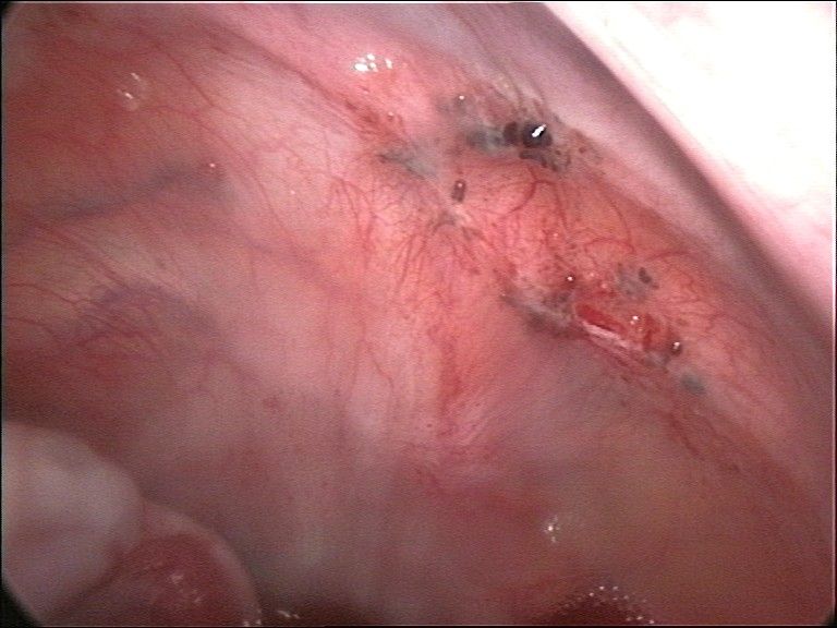
Laparoscopy is similar to hysteroscopy, but a camera is inserted into the abdomen, allowing for visualisation of the external surfaces of abdominal and pelvic viscera which can be helpful in diagnosing conditions like endometriosis.
Hysteroscopies and laparoscopies both have the benefit of being able to be therapeutic (i.e. you can treat the condition there and then) as well as being diagnostic.
The overall goal in the management of menstrual disorders is to manage the underlying condition. It is very important in gynaecology to create a patient-centred management plan. The mainstay of pharmacological management is the use of hormones such as gonadotrophins, progesterone, combined oral contraceptive pill, or hormone replacement therapy. Symptoms can be controlled with supportive measures like analgesia. Depending on the condition, a surgical approach may be needed.
It is also important to consider the patient’s fertility as this will affect whether contraceptive options are appropriate for the patient – is this patient considering pregnancy at the moment? Are they likely to want a pregnancy soon?

Image - Endometrial tissue in the peritoneum causing endometriosis as seen on laparoscopy
Public Domain Source by Hic et nunc [Public domain]
Edited by: Dr. Maddie Swannack
Reviewed by: Dr. Thomas Burnell
- 3339

