Next Lesson - Atherosclerosis
Abstract
- If the blood, blood flow, or vessel wall is abnormal or disrupted, platelets can aggregate resulting in the pathological formation of a blood clot – a thrombus.
- This thrombus can remain in its site of origin, or it can travel to distant parts of the body and block a blood vessel (at which point it becomes an embolus).
- Thrombi and emboli can cause infarction and ischaemia, damaging the affected tissues.
- Sudden occlusion of a limb artery results in the six P's: pain, pulselessness, parasthesia, pallor, paralysis and perishing cold.
- Thrombus formation in the veins will cause an acute inflammatory response resulting in the five cardinal signs: rubor, calor, tumor, dolor and loss of function. Embolisation can lead to a pulmonary embolism.
Core
A thrombus is the name given to a solid mass which is formed from the constituents of the blood within the heart or vessels during life. Thrombosis is the process of forming this thrombus and it occurs when the normal haemostatic mechanisms are turned on inappropriately.
There are three main factors which will increase the likelihood of thrombosis occurring. Collectively these are known as Virchow’s Triad of thrombosis and consist of the following:
- Changes in the vascular wall (endothelial damage)
- E.g. After myocardial infarction, surgery, chemical damage or ruptured atherosclerotic plaque
- Changes in the blood flow (slow or turbulent flow)
- E.g. Due to long periods of immobility, varicose veins or venous obstruction
- Changes in the blood (hypercoagulability)
- E.g. Due to cancer, thrombophilia, inflammatory disease, smoking or pregnancy
Thrombosis occurs via the same mechanisms as in haemostasis, i.e. platelets are activated and bound together by fibrin, which goes on to trap red blood cells to form a clot.
Thrombi are laminated structures (they are made up of layers), and the lines of lamination are known as lines of Zahn. This is a consequence of the way thrombi are formed.
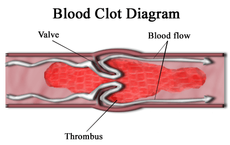
As platelets are the smallest constituents of blood, they tend to be pushed by the force of blood flow to the walls of the blood vessel and so are more concentrated along the endothelium. The platelets are therefore more likely to catch in any crevices, such as behind the valves of veins or abnormalities in the vessel wall. These caught platelets can form an aggregate and stick to the wall of the vessel. As blood continues to flow, additional platelets can join the aggregate, forming a white layer of platelets. Red blood cells then get caught in the aggregate, creating a red layer, followed by another white layer of platelets and the process repeats. These continual additions will result in the laminar form of the thrombus.

Diagram - The formation of a blood clot in a vein around a valve. Blood flow through the vein is reduced due to the obstruction.
Creative commons source by Persian Poet Gal [CC BY-SA 3.0 (http://creativecommons.org/licenses/by-sa/3.0/)]
Lines of Zahn are more visible in arterial thrombi as blood flows directly over the surface of the forming thrombi in arteries, whereas there is a degree of shielding by the valves in veins. Clots formed post-mortem have no lines of Zahn as there is no blood flow.
There are several outcomes which can arise from thrombus formation:
- Lysis (breakdown) of the thrombus.
- This is the most favourable outcome. Blood flow is re-established completely in this case. It is most likely when thrombi are small.
- Propagation
- The thrombus will progressively spread. It can become bigger, and form new thrombi downstream of the original.
- Organisation
- Fibroblasts act on the thrombus and scar tissue forms where the thrombus is. In this case the lumen of the blood vessel will remain partially occluded due to the scar.
- Recanalisation
- One or more channels can be formed through the thrombus like tunnels, so blood flow is re-established but usually incompletely.
- Embolism
- Part of the thrombus breaks off and travels through the bloodstream to lodge at a distant site.
Embolism is the blockage of a blood vessel by solid, liquid or gas at a site distant from its origin. 90% of emboli are thromboemboli (originating from a thrombus), and most thromboemboli arise in the deep veins of the legs - called deep vein thrombosis (DVT).
Emboli carried in the veins will most often travel to the right side of the heart and enter the pulmonary circulation to block an artery in the lungs. This is called a pulmonary embolism.
In arteries, the blood flows from larger vessels to smaller vessels, so emboli carried in the arteries will lodge in smaller arteries downstream. Emboli from systemic arteries or the left side of the heart can impact themselves in the systemic circulation. If this occurs in the brain it can cause a stroke.
There are other types of embolism apart from thromboemboli that are rarer. These include:
- Fat and bone marrow embolism
- Can occur after breaking a bone, especially a large one such as the femur. The fat or bone marrow form an insoluble embolism which travels through the blood and blocks a vessel.
- Gas embolism
- Air embolism can occur following physical trauma: veins can draw in air from the external environment. Air is transported to the right side of the heart and create a frothy mass that blocks circulation.
- ‘The bends’ are another process of forming a gas embolism. If a diver surfaces from deep water too quickly, the sudden drop in pressure can cause dissolved gases in the blood to come out of solution and form bubbles in the circulation.
- Amniotic fluid embolism
- A rare but dangerous complication of labour and C-section where amniotic fluid enters the maternal circulation through a tear in the amniotic membranes.
- Talcum embolism
- Microscopic particles can enter the circulation of IV drug users.
For more information on artery and vein diseases, click here for the article in the cardiovascular system module. A summary of the clinical effects of arterial and venous thrombosis and embolism is below.
Examples of how this can happen include: an abdominal aortic aneurysm (AAA), atherosclerosis, or clot formation in the left atrium in atrial fibrillation. In each of these examples one or more elements of Virchow's triad is affected.
Sudden occlusion of an artery is very serious and needs to be treated quickly to avoid damage to the limb or organ.
The signs and symptoms of occlusion of an artery the a limb are the six P’s, and result from the lack of blood perfusing the tissues:
- Pain
- Paralysis
- Paraesthesia
- Pallor
- Perishingly cold to touch
- Pulseless
Thrombus formation in veins occurs slower than in arteries, giving time for the body to mount an acute inflammatory response. Therefore the signs of thrombus formation in veins are the five cardinal signs of inflammation: rubor, calor, tumor, dolor and loss of function.
The most common site for thrombus formation is in the deep veins of the legs. This is commonly due to immobility. As the calf muscles contract during movement, the venous blood is pushed back towards the heart. Therefore by not moving, for example in a long haul flight, the blood in the veins doesn’t flow as much, causing stasis of blood in the legs. This is one of Virchow’s triad, and a clot may develop. This is called a deep vein thrombosis (DVT). The five signs of inflammation may be seen in the calf.
If the DVT embolises, the fragment will travel through the venous system towards the right side of the heart and enter the pulmonary circulation. It will then lodge in an artery, causing a pulmonary embolism.
Edited by: Bethany Turner
- 8631

